Building Confidence One Smile at a Time.

Tonsil Stone or Something More Serious? How to Identify It, Treat It & Know When to Act
Image:
Severity:
Teeth Problems:
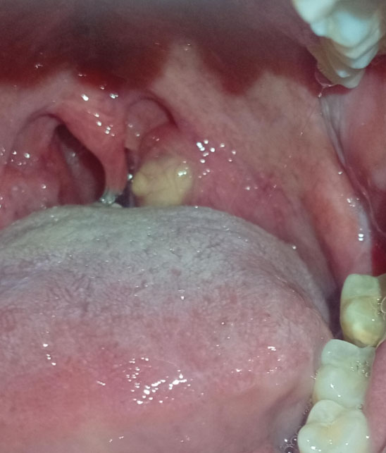
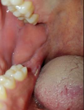
I enlarged the uploaded image to 200% for visual inspection and focused on the tonsillar area, back teeth, tongue, and surrounding soft tissue.
Main finding: Could this be a tonsil stone?
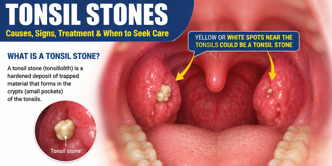
Tonsil stones commonly appear as white or yellow deposits lodged in tonsillar crypts and can be associated with bad breath, bad taste, throat irritation, cough, or the sensation that something is stuck in the throat.
100% visual assessment
- The visible yellowish/cream mass is the most important finding.
- Its location appears consistent with the region where a tonsil stone could become trapped.
- There is surrounding pink/red tissue that may indicate local irritation or inflammation.
- I don't see enough evidence in this photograph to call it a peritonsillar abscess.
Area 2 — Teeth
- The visible posterior teeth show some yellowish discoloration/deposits.
- This photograph does not provide enough coverage to diagnose the full condition of all teeth.
- A clinical dental examination is needed for cavities, calculus, periodontal pockets and bone support.
Area 3 — Tongue
- The tongue has a noticeable coating toward the posterior portion.
- Tongue coating can contribute to oral odor and bacterial accumulation.
- It can also coexist with tonsil stones.
What else could the yellow area be?
The differential includes:
- Tonsil stone — possible/likely
- Food debris trapped in a tonsillar crypt
- Tonsillar inflammation with exudate
- Less commonly, another lesion or abnormal tissue
A clinician can inspect the tonsil directly and determine whether the material is actually embedded in a tonsillar crypt. Tonsil stones can sometimes be diagnosed simply by oral/throat examination.
What should happen next?
Step 1 — Dental/oral examination
- Examine posterior teeth and gums.
- Check plaque/calculus.
- Check for cavities and periodontal disease.
- Clean the teeth professionally if indicated.
Step 2 — Tonsil examination
A dentist, physician or ENT specialist should inspect the suspected area.
Step 3 — Determine whether it is actually a stone
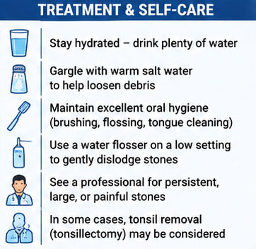
If confirmed as a tonsil stone, treatment may range from observation and oral hygiene to professional removal when symptomatic or recurrent.
Step 4 — Look for the underlying cause
Repeated tonsil stones can be associated with deeper tonsillar crypts and previous tonsil inflammation/infections.
If you give it 14 days
If this is a small uncomplicated tonsil stone, it may loosen or fall out by itself. Warm salt-water gargling and good oral hygiene can help; some stones require professional removal.
But 14 days should not be treated as a guaranteed healing period.
If the underlying problem is tonsillitis, a persistent lesion, or another infection, simply waiting 14 days could allow the problem to continue.
What could "scale up" if ignored?
Watch for:
Tonsil stone → persistent irritation → recurrent tonsil inflammation/infection → increasing discomfort, bad breath or swallowing problems.
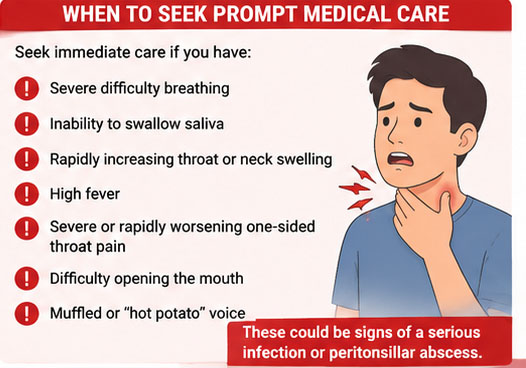
More importantly, if the area is actually an infection rather than a stone, progression can be more serious. A peritonsillar abscess can cause severe one-sided throat pain, difficulty swallowing, difficulty opening the mouth, fever, swelling and a muffled voice.
Emergency warning: difficulty breathing, inability to swallow saliva, rapidly increasing throat/neck swelling, or severe worsening symptoms should be evaluated urgently rather than waiting 14 days.
My visual conclusion
Suspected tonsil stone: YES — plausible from the image.
Confirmed tonsil stone: NO — requires direct clinical examination.
Visible local irritation: POSSIBLE.
Definite tonsillitis: Cannot determine from this photograph.
Definite abscess: Not established from this image.
I would recommend having the suspected tonsil stone and the posterior teeth examined during the same visit, because both oral hygiene and tonsillar crypts can contribute to persistent bad breath.
For a suspected tonsil stone that keeps returning, an ENT (ear, nose and throat) evaluation may also be appropriate in addition to the dental examination.
Frequently Asked Questions About Tonsil Stones
1. Are tonsil stones dangerous?
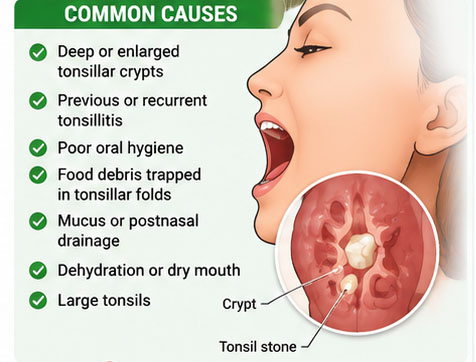
Most tonsil stones are harmless and do not require medical treatment. They form when food particles, bacteria, mucus, and dead cells become trapped in small pockets in the tonsils and gradually harden. However, persistent pain, bleeding, swelling, or difficulty swallowing should not automatically be attributed to tonsil stones. Consult a healthcare professional if these symptoms occur.
2. How can I tell if I have tonsil stones or a more serious condition?
Tonsil stones often appear as small white or yellow lumps on the tonsils. They may cause bad breath, an unpleasant taste, or a sensation that something is stuck in the throat.
Other conditions, including tonsillitis, throat infections, and occasionally more serious diseases, can cause similar symptoms. A high fever, severe or worsening throat pain, significant swelling, or persistent one-sided symptoms deserve medical assessment. You cannot reliably diagnose every throat condition by appearance alone.
3. Can tonsil stones be a sign of cancer?
Tonsil stones themselves are not cancer. However, a persistent lump, an unexplained enlarged tonsil on one side, a neck lump, blood in saliva, or ongoing difficulty swallowing should be evaluated by a doctor. These symptoms can have several causes and do not automatically mean cancer, but they should not be ignored.
4. Why do my tonsil stones smell so bad?
Tonsil stones can develop an unpleasant smell because bacteria and trapped debris accumulate in the tonsil pockets. These bacteria can produce sulfur-containing compounds that contribute to bad breath. Brushing your teeth, flossing, cleaning your tongue, drinking enough water, and gargling with warm salt water may help reduce the problem.
5. How can I remove tonsil stones safely at home?
Start with gentle measures, such as gargling with warm salt water, maintaining good oral hygiene, and drinking sufficient water. Some stones come out naturally when swallowing or coughing.
Avoid sharp objects, fingernails, forceful squeezing, and high-pressure water jets directed at the tonsils. These can injure delicate tissue and cause bleeding or infection. If a stone is deeply lodged, painful, or difficult to remove, ask a healthcare professional for assistance.
6. Can tonsil stones go away on their own?
Yes. Many tonsil stones loosen and fall out naturally without treatment. Small stones that do not cause discomfort can often be left alone. Seek medical advice if they persist, repeatedly return, or cause pain, swelling, or swallowing problems.
7. Why do I keep getting tonsil stones even when I brush my teeth?
Tonsil stones can recur because some people naturally have deeper pockets or folds in their tonsils. Previous tonsil infections may also contribute to irregularities where debris becomes trapped. Good oral hygiene helps reduce debris and bacteria, but it cannot always prevent recurrence.
8. Can tonsil stones cause bad breath even if I brush and floss daily?
Yes. Tonsil stones can contribute to bad breath even when your teeth and gums are clean. However, bad breath can also come from gum disease, tooth decay, dry mouth, sinus problems, or other conditions. If the odor continues despite good oral hygiene, consider a dental examination and, when appropriate, a medical assessment.
9. Do I need antibiotics to treat tonsil stones?
Usually, no. Tonsil stones alone do not normally require antibiotics. Antibiotics treat certain bacterial infections, not the hardened debris that forms the stones. Do not take leftover antibiotics or start them without medical advice. If you develop fever, worsening throat pain, or other signs of infection, seek medical assessment.
10. When should I see a doctor or an ENT specialist?
Arrange a medical appointment if you have recurring or troublesome tonsil stones, persistent sore throat, unexplained one-sided tonsil enlargement, bleeding, ongoing ear pain, a neck lump, or difficulty swallowing.
Seek emergency care immediately if you have difficulty breathing, cannot swallow your saliva, or develop rapidly increasing throat or neck swelling. Severe difficulty swallowing or breathing should never be treated as an ordinary tonsil stone problem.
11. Can tonsil stones cause ear pain or the feeling that something is stuck in my throat?
Yes. Tonsil stones may cause referred ear discomfort or a sensation of something lodged in the throat. However, ear pain and persistent throat discomfort can have other causes. If symptoms are severe, last more than a few days, worsen, or occur with fever or difficulty swallowing, seek medical advice rather than assuming a tonsil stone is responsible.
12. Should I have my tonsils removed if tonsil stones keep coming back?
Not necessarily. Most people with tonsil stones do not need surgery. If stones repeatedly cause significant discomfort, persistent bad breath, or other problems despite appropriate care, an ENT specialist can discuss available options. Tonsillectomy, which removes the tonsils, may be considered in selected cases after discussing the benefits, risks, and recovery time.
13. Are tonsil stones contagious?
No. Tonsil stones themselves are not contagious. However, some infections that affect the tonsils or throat can spread between people. If you have fever, a significant sore throat, or other symptoms of an infection, seek appropriate medical advice.
14. Can drinking water and gargling with salt water prevent tonsil stones?
Useful information
1. Gum Swelling Near Back Teeth
Best for readers with swelling near a molar or back tooth.
Read the related case study
2. Severe Tartar Buildup and Gum Inflammation
For readers concerned about bleeding, plaque, tartar, or red gums.
Explore gum inflammation case studies
3. Dental Abscess and Oral Infection
For readers who notice pus, a bad taste, tooth pain, or increasing swelling.
Explore oral infection guides
These measures may help reduce debris and ease mild irritation, but they cannot guarantee that stones will not return. Brush and floss regularly, gently clean your tongue, stay hydrated, and gargle with warm salt water if appropriate. Avoid aggressive attempts to clean inside the tonsils.
Important Health Advice
Tonsil stones are usually harmless, but not every white spot, lump, or sore area at the back of the throat is a tonsil stone. If you are unsure what you are seeing, have persistent symptoms, or notice unusual swelling or bleeding, arrange an examination with a qualified healthcare professional.
This article provides general educational information and does not replace an individual medical examination or diagnosis.
Related Clinical Cases
Patients with lower front gum inflammation may also experience tartar buildup, periodontal disease, gum abscesses, or tooth decay. Explore these related clinical case studies.



Other topics:
Other Dental Clinic