The Hidden Threat: What Back Tooth Pain and Bleeding Gums Could Mean for Your Health

Severity:
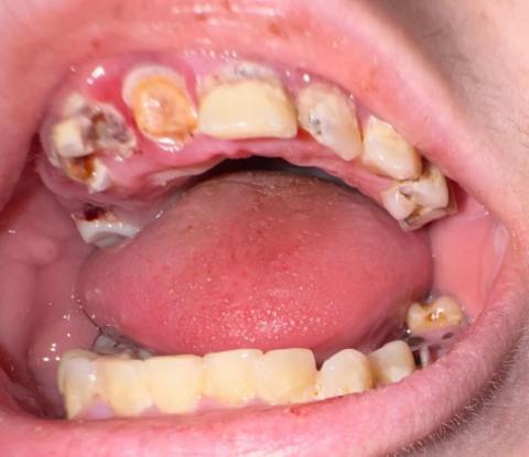
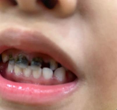
Teeth Problems:
Deep Examination & Analysis
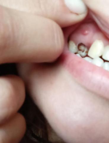
Based on the image provided, there are several significant dental health concerns that require immediate professional attention:
-
Gross Caries (Severe Decay): The lower molar (bottom right of the photo) shows a large, dark cavitation covering most of the occlusal (biting) surface. This indicates extensive tooth structure loss.
-
Likely Pulp Exposure: Given the depth of the dark area on the lower molar, the decay has likely reached the pulp (nerve) of the tooth.