Building Confidence One Smile at a Time.

Bone Density Evaluation: DEXA Scan Explained — A U.S. Dentist’s Guide to Bone Health, Osteoporosis, and Dental Implant Planning
image hero:
As a dentist practicing in the United States, I routinely evaluate patients who are concerned about missing teeth, jawbone loss, dentures, periodontal disease, and dental implants. One issue that deserves much more attention is overall bone health.
Patients sometimes assume that because a dental X-ray shows adequate bone in the jaw, their skeletal system is healthy. That conclusion is not necessarily correct.
Bone density is a systemic health issue. Osteoporosis can weaken bones throughout the body and increase the risk of fractures, while local jawbone conditions can simultaneously affect tooth support and dental implant planning.
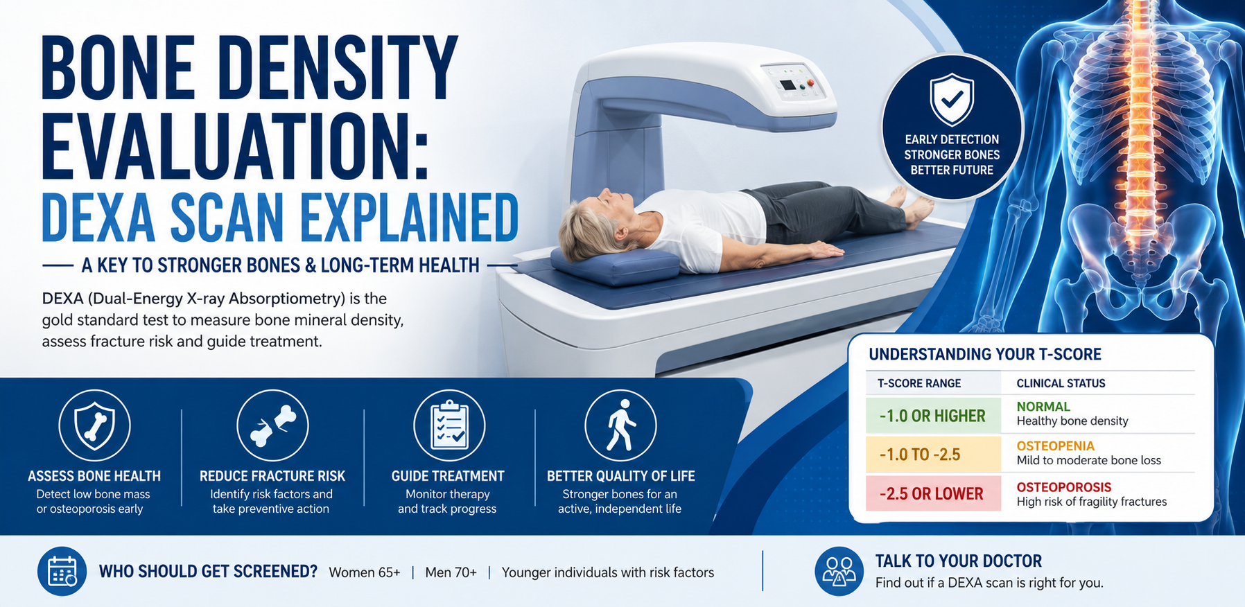
One of the most important tools for evaluating systemic bone mineral density is the DEXA or DXA scan, short for dual-energy X-ray absorptiometry.
A DXA scan provides a quantitative measurement of bone mineral density and is commonly used to identify osteoporosis, low bone mass, and fracture risk.
From a dental perspective, understanding a patient's bone health is particularly important when discussing tooth loss, periodontal disease, implant treatment, oral surgery, and medications used to treat osteoporosis.
How Common Is Osteoporosis in the United States?
The burden of osteoporosis in the United States is significant.
According to CDC data from the National Health and Nutrition Examination Survey (NHANES), approximately 12.6% of U.S. adults age 50 and older had osteoporosis based on bone density measurements at the femoral neck or lumbar spine during 2017–2018.
The prevalence was substantially higher among women:
- Women: 19.6%
- Men: 4.4%
- Adults overall: 12.6%
In addition, approximately 43.1% of adults age 50 and older had low bone mass, sometimes referred to clinically as osteopenia depending on the patient's circumstances and diagnostic criteria.
Low bone mass affected approximately:
- 51.5% of women
- 33.5% of men
The CDC also reported that osteoporosis prevalence among women increased from approximately 14.0% in 2007–2008 to 19.6% in 2017–2018. In contrast, the prevalence among men did not show a statistically significant change during those survey periods, and overall low-bone-mass prevalence remained relatively stable.
These numbers demonstrate why bone health should not be viewed as an exclusively orthopedic or geriatric concern.
What Is Bone Density?
Bone is living tissue.
Throughout life, old bone is continuously removed and replaced with new bone. During childhood and adolescence, bone formation exceeds bone breakdown, allowing the skeleton to grow and become stronger.
Peak bone mass is generally achieved by the mid- to late 20s. After that point, maintaining bone strength becomes increasingly important because the balance between bone formation and bone resorption can change with aging.
Several factors influence bone density, including:
- Age
- Genetics
- Sex
- Menopause
- Physical activity
- Calcium intake
- Vitamin D status
- Protein intake
- Smoking
- Alcohol consumption
- Certain medications
- Certain medical conditions
- Previous fractures
- Body weight
Bone loss can occur silently.
That is one reason osteoporosis is sometimes called a silent disease. A person may feel completely healthy until a fracture occurs.
What Is a DEXA or DXA Scan?
DEXA and DXA refer to the same basic technology:
Dual-Energy X-ray Absorptiometry.
The examination uses very low-dose X-rays to measure bone mineral density.
The most clinically important measurements generally involve areas such as the:
- Hip
- Femoral neck
- Lumbar spine
The test is noninvasive and usually takes only a short period of time.
Unlike a conventional dental X-ray, which provides localized images of teeth and jaw structures, a DXA examination is designed specifically to quantify bone mineral density at clinically important skeletal sites.
The Bone Health and Osteoporosis Foundation describes DXA as the most common and versatile bone-density test and notes that it can help diagnose osteoporosis before a fracture occurs, estimate future fracture risk, and monitor treatment.
Why Would a Dentist Care About a Patient's Bone Density?
This is an important question.
Dentists do not diagnose osteoporosis simply by looking at a patient's teeth.
However, dentists frequently encounter patients who may have risk factors for skeletal bone loss.
For example, a patient may present with:
- Multiple missing teeth
- Advanced periodontal disease
- Tooth mobility
- Denture-related problems
- Severe alveolar bone loss
- A history of fractures
- Long-term medication use
- Planned tooth extraction
- Planned dental implant treatment
- Treatment with osteoporosis medications
The dentist's role is to recognize when the patient's overall medical history may have implications for oral treatment and communicate appropriately with the patient's physician.
A dental X-ray should not be considered a substitute for a medical DXA scan.
Bone Density and the Jaw Are Not the Same Thing
This distinction is extremely important.
A DXA scan evaluates bone mineral density at specific skeletal sites, particularly the hip and spine.
Dental imaging evaluates the jaws and teeth.
A patient may have reduced skeletal bone density without having obvious dental symptoms.
Conversely, significant jawbone loss may result from periodontal disease, tooth loss, infection, trauma, or other local conditions rather than systemic osteoporosis alone.
This means a dentist should avoid telling a patient:
"Your jawbone looks good, so your bone density must be normal."
That conclusion cannot be made from a routine dental examination.
Similarly, a diagnosis of osteoporosis does not automatically mean that dental implants are impossible.
Implant treatment requires a comprehensive evaluation of local bone anatomy, oral health, systemic health, medications, and individual risk factors.
Understanding the DEXA T-Score
One of the most important numbers on a DXA report is the T-score.
For postmenopausal women and men age 50 and older, T-scores are commonly interpreted using the following categories:
| T-Score | Classification |
|---|---|
| -1.0 or higher | Normal bone density |
| Between -1.0 and -2.5 | Low bone mass / osteopenia |
| -2.5 or lower | Osteoporosis |
The T-score compares the patient's bone mineral density with that of a healthy young-adult reference population.
A lower number indicates lower bone mineral density.
For example:
T-score: -0.5
Generally falls within the normal range.
T-score: -1.7
Falls within the low-bone-mass range.
T-score: -2.8
Falls within the osteoporosis range.
However, a DXA result should never be interpreted in isolation. The patient's age, sex, fracture history, medical conditions, medications, and overall fracture risk also matter.
T-Score vs. Z-Score
Patients sometimes see both a T-score and a Z-score on their DXA report.
They are not interchangeable.
T-Score
The T-score compares bone density with a young-adult reference population and is commonly used for osteoporosis classification in postmenopausal women and men age 50 and older.
Z-Score
The Z-score compares bone density with an age- and sex-matched reference population.
Z-scores are particularly useful in younger individuals.
The Bone Health and Osteoporosis Foundation notes that a Z-score of -2.0 or lower is considered below the expected range for age and should prompt consideration of the patient's medical history and possible secondary causes of bone loss.
This distinction is important because the same numerical bone-density value can have different clinical implications depending on the patient's age and reproductive status.
Who Should Consider a Bone Density Evaluation?
Screening recommendations can vary depending on the organization and the patient's individual risk profile.
The current U.S. Preventive Services Task Force recommendation states that women 65 years and older should be screened for osteoporosis to prevent fractures.
The USPSTF also recommends screening postmenopausal women younger than 65 who have increased fracture risk based on clinical risk assessment.
For men, the USPSTF currently states that evidence is insufficient to determine the overall benefits and harms of population-based screening. This is important because it does not mean men cannot or should not receive DXA testing when clinically appropriate. Physicians may use clinical judgment based on individual risk factors.
Other professional organizations use different approaches. For example, the Bone Health and Osteoporosis Foundation advises patients to discuss bone-density testing with their healthcare provider if they are a woman age 65 or older, a man age 70 or older, or have experienced a fracture after age 50.
Therefore, screening should be individualized rather than based solely on age.
Risk Factors That May Lead to Earlier Evaluation
A patient may require evaluation before the traditional screening age when significant risk factors are present.
Important risk factors can include:
- Previous fragility fracture
- Low body weight
- Family history of hip fracture
- Smoking
- Excessive alcohol consumption
- Menopause
- Long-term corticosteroid use
- Certain endocrine disorders
- Certain gastrointestinal disorders
- Some cancer treatments
- Reduced physical activity
- Certain medications associated with bone loss
The USPSTF specifically identifies menopausal status, low body weight, parental history of hip fracture, cigarette smoking, and excess alcohol consumption among important risk factors considered when evaluating postmenopausal women younger than 65.
Why Physical Activity Matters for Bone Health
Bone responds to mechanical loading.
Weight-bearing activity and resistance exercise can help support skeletal health, while strength and balance training can also reduce fall risk.
Examples include:
- Walking
- Stair climbing
- Resistance training
- Strength exercises
- Appropriate balance exercises
However, people who already have osteoporosis should not simply begin high-impact exercise without medical guidance.
Exercise should be matched to the individual's fracture risk and physical condition.
The National Institute of Arthritis and Musculoskeletal and Skin Diseases recommends strength or resistance exercise and emphasizes safe, individualized activity for people with osteoporosis.
Calcium, Vitamin D, and Bone Health
Nutrition is another major component of skeletal health.
Calcium is an essential mineral for bones, while vitamin D helps the body absorb calcium.
The NIH notes that inadequate calcium intake can cause the body to draw calcium from bone stores over time, potentially weakening bones.
General recommended calcium intake varies by age and sex.
For example, NIH guidance lists approximately:
- Adults 19–50: 1,000 mg/day
- Women over 50: 1,200 mg/day
- Men 51–70: 1,000 mg/day
- Men over 70: 1,200 mg/day
Vitamin D requirements also vary by age.
Importantly, patients should not automatically begin high-dose supplements simply because they are concerned about osteoporosis. Dietary intake, medical history, kidney function, medications, and laboratory findings may influence the appropriate approach.
Bone Health and Dental Implants
This is where the subject becomes especially relevant to dentistry.
Dental implants require sufficient local bone volume and quality for predictable placement and long-term function.
However, systemic osteoporosis does not automatically rule out dental implant treatment.
The dentist must evaluate the actual implant site using appropriate dental imaging, clinical examination, periodontal evaluation, and treatment planning.
Depending on the case, three-dimensional imaging such as cone-beam computed tomography may be used to evaluate:
- Bone volume
- Ridge width
- Ridge height
- Anatomical structures
- Sinus position
- Inferior alveolar nerve location
- Potential implant positioning
A medical DXA scan answers a different question.
It evaluates systemic bone mineral density rather than providing the detailed local anatomical information needed to place a dental implant.
Therefore:
DXA = systemic bone-density assessment
Dental imaging = local jaw and implant-site assessment
The two examinations can complement one another but should not be confused.
Osteoporosis Medications and Dental Treatment
Another important issue for dentists is the medication history.
Some patients with osteoporosis are treated with antiresorptive medications, including bisphosphonates or denosumab.
These medications can reduce fracture risk and are important treatments for appropriately selected patients.
However, dentists need to know when patients are taking these medications because of the rare complication known as:
Medication-Related Osteonecrosis of the Jaw (MRONJ).
The American Dental Association describes MRONJ as a rare but serious adverse effect associated with antiresorptive medications. Risk varies according to the medication, dosage, duration, underlying disease, and other patient factors.
Importantly, patients should not stop osteoporosis medication on their own because they need dental treatment.
The ADA emphasizes that the fracture-prevention benefits of antiresorptive therapy generally outweigh the low MRONJ risk in patients receiving these medications for osteoporosis.
Dental and medical providers should coordinate treatment when appropriate.
Does Osteoporosis Cause Tooth Loss?
Osteoporosis and tooth loss are related to overall bone health, but it would be inaccurate to say:
"Osteoporosis causes your teeth to fall out."
Tooth loss has many potential causes.
The most important dental causes include:
- Periodontal disease
- Dental caries
- Tooth fracture
- Infection
- Trauma
- Poor oral hygiene
- Failed restorations
- Other local dental conditions
Periodontal disease can destroy the bone supporting individual teeth.
That process is different from the systemic bone loss measured by a DXA scan.
A patient can have excellent systemic bone density and still lose teeth from severe periodontal disease.
Likewise, a patient with osteoporosis may have healthy natural teeth.
Why Dentists Should Ask About Fracture History
A fracture after relatively minor trauma can be an important medical clue.
For example, an older adult who experiences a fracture after a simple fall may require evaluation for osteoporosis.
From the dental perspective, this information can become relevant when reviewing the patient's medical history before:
- Extractions
- Periodontal surgery
- Implant placement
- Bone grafting
- Oral surgery
- Denture treatment
A dentist may recognize a potential systemic health issue and recommend that the patient discuss bone-density evaluation with their physician.
This is one of the advantages of comprehensive dental care: the mouth can sometimes provide an opportunity to identify broader health concerns.
What Happens During a DXA Examination?
A typical DXA examination is straightforward.
The patient lies on a specialized scanning table while the machine measures bone mineral density at selected skeletal sites.
The examination uses a low amount of radiation.
The results are analyzed to determine bone mineral density and generate measurements such as the T-score and Z-score.
The physician then interprets those results alongside the patient's clinical history.
A DXA scan is not a painful procedure.
It does not involve needles or surgical intervention.
The exact preparation requirements can vary by facility, so patients should follow the instructions provided by the imaging center.
What Happens After an Abnormal DEXA Result?
An abnormal result does not mean that treatment is automatically the same for every patient.
The physician may evaluate:
- Fracture history
- Age
- Sex
- Menopausal status
- Family history
- Medication history
- Calcium and vitamin D intake
- Physical activity
- Smoking
- Alcohol consumption
- Other medical conditions
- Additional laboratory testing when appropriate
- Overall fracture risk
Some patients may require lifestyle intervention alone.
Others may benefit from pharmacologic treatment.
Patients with very low bone density or previous fragility fractures may require more intensive management.
The important point is that a DXA result is a starting point for clinical decision-making, not the entire diagnosis.
Bone Density Evaluation Is About Prevention
The greatest value of bone-density evaluation is not simply giving a patient a number.
The real goal is prevention.
If osteoporosis is identified before a serious fracture occurs, the patient and healthcare team have an opportunity to address modifiable risk factors and, when appropriate, use evidence-based treatment to reduce fracture risk.
The USPSTF found that screening higher-risk women age 65 and older can provide a small absolute reduction in hip and major osteoporotic fractures, supporting the value of appropriate screening.
That makes early evaluation particularly important as the U.S. population ages.
A Dentist's Perspective: Don't Look at Bone Health in Isolation
When I evaluate a patient for dental treatment, I am not looking only at the teeth.
I want to understand the patient's overall health.
For an older adult with multiple missing teeth, periodontal disease, a history of fractures, osteoporosis treatment, or planned implant surgery, the medical history becomes especially important.
A dental examination can identify local bone conditions.
A dental CT can show the three-dimensional anatomy of the jaw.
A DXA scan can evaluate systemic bone mineral density.
A physician can evaluate the patient's overall fracture risk and determine whether medical treatment is necessary.
These evaluations answer different questions.
Together, they provide a more complete picture.
Frequently Asked Questions
Is a DEXA scan the same as a dental X-ray?
No.
A DXA scan is specifically designed to measure bone mineral density at skeletal sites such as the hip and spine.
A dental X-ray evaluates teeth and structures of the jaws.
They serve different purposes.
Can a dentist order a DEXA scan?
This depends on the dentist's scope of practice, state regulations, healthcare system, and individual clinical circumstances. In many situations, the dentist will recommend that the patient discuss bone-density testing with their physician or other medical provider.
Does osteoporosis mean I cannot get dental implants?
Not necessarily.
Implant candidacy depends on local jawbone anatomy, oral health, systemic health, medications, smoking status, healing potential, and other factors.
Osteoporosis alone does not automatically mean that implants are contraindicated.
Can dental X-rays diagnose osteoporosis?
No.
Dental radiographs may show changes in jawbone appearance, but they are not a replacement for a medical DXA examination.
Is osteopenia the same as osteoporosis?
No.
Osteopenia generally describes bone density that is below the normal range but not low enough to meet the conventional DXA threshold for osteoporosis.
However, fracture risk depends on more than the T-score alone.
Should men have their bone density checked?
Men can develop osteoporosis.
The current USPSTF states that evidence is insufficient to recommend for or against routine population screening in men, but clinicians may use individual risk factors and clinical judgment to determine whether testing is appropriate.
Should I stop osteoporosis medication before dental treatment?
Do not stop prescribed osteoporosis medication on your own.
Some osteoporosis medications are important for preventing serious fractures. Dental treatment should be coordinated with the patient's prescribing physician when medication management becomes relevant.
The Bottom Line
Bone density is an important component of healthy aging.
Current U.S. data show that osteoporosis affects approximately 12.6% of adults age 50 and older, while another 43.1% have low bone mass. Women carry a substantially greater burden of osteoporosis, and the prevalence among women increased during the CDC's 2007–08 to 2017–18 survey period.
A DXA/DEXA scan is one of the principal tools used to measure systemic bone mineral density.
From a dental perspective, understanding bone health is particularly important when treating older adults, patients with a history of fractures, patients with osteoporosis, and patients taking antiresorptive medications.
But remember that systemic bone density and jawbone health are not identical.
A comprehensive approach may involve:
Medical history → fracture-risk assessment → DXA when indicated → dental examination → periodontal evaluation → appropriate dental imaging → individualized treatment planning.
If you are concerned about osteoporosis, have experienced a fracture after a minor injury, or have risk factors for bone loss, discuss bone-density evaluation with your physician.
And if you are considering dental implants or oral surgery while taking osteoporosis medication, make sure your dentist knows exactly which medication you take, the dose, and how long you have been taking it.
Healthy bones support mobility, independence, and quality of life. Good oral health is another essential part of healthy aging.
Related Clinical Cases
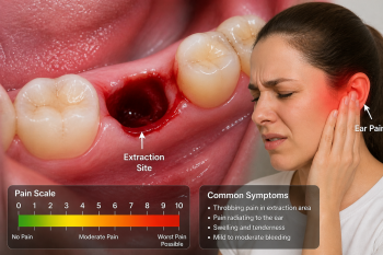
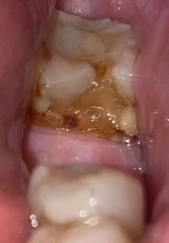
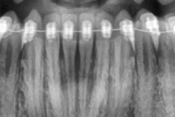
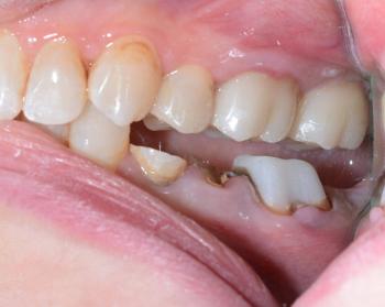
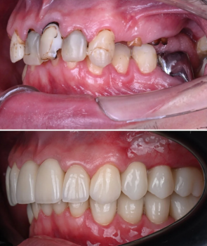
Patients with lower front gum inflammation may also experience tartar buildup, periodontal disease, gum abscesses, or tooth decay. Explore these related clinical case studies.



Other topics:
Other Dental Clinic
 |
 |
 |
 |





