Healthy Teeth, Happy Life.

Bone Loss After Tooth Extraction: A German Dentist’s Clinical Review, Case Analysis, and Expert Advice
image hero:
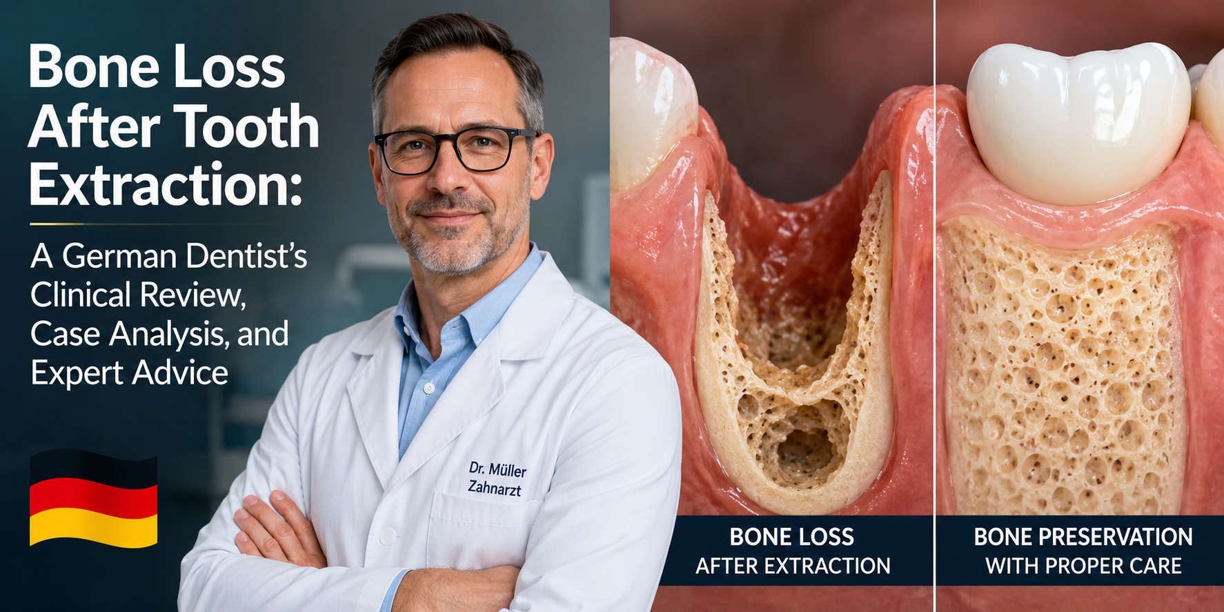
Tooth extraction removes the tooth, but it does not stop the biological changes that occur inside the jaw.
One of the most important consequences is alveolar bone loss, also called post-extraction ridge resorption. The bone that previously surrounded and supported the tooth begins to remodel because the tooth root and the mechanical stimulation associated with it are no longer present.
From a German evidence-based dentistry perspective, this is an important issue to discuss with every patient who has lost a tooth, particularly when a dental implant may be considered in the future.
Scientific reviews consistently show that the extraction socket undergoes measurable reduction in both width and height, with horizontal loss often greater than vertical loss. One systematic review reported an average reduction in ridge width of approximately 3.87 mm and an average mid-buccal height loss of approximately 1.67 mm.
1. What Happens to the Bone After Extraction?
A tooth is not simply sitting inside the jawbone.
Its root is surrounded by the periodontal ligament, which connects the tooth to the alveolar bone. The surrounding bone is constantly responding to mechanical forces generated during chewing.
After extraction, that biological environment changes.
The body begins a normal healing process:
Extraction → blood clot → granulation tissue → new bone formation → bone remodeling → ridge contraction
This process is not necessarily a disease.
It is a natural biological response.
The problem is that the healed ridge can become significantly narrower and sometimes shorter than the original tooth-supporting bone.
2. How Much Bone Can Be Lost?
The amount varies considerably between patients.
Factors include:
-
Tooth location
-
Size and shape of the extraction socket
-
Thickness of the facial/buccal bone
-
Presence of periodontal disease
-
Infection around the tooth
-
Smoking
-
Surgical trauma
-
Individual healing characteristics
-
Systemic factors affecting bone metabolism
-
Whether ridge-preservation treatment was performed
Research shows that extraction alone commonly results in measurable dimensional changes.
A systematic review found that natural healing after extraction can produce approximately 2.6–4.6 mm of horizontal reduction and approximately 0.4–3.9 mm of vertical reduction, depending on the study and clinical situation.
This is why I would not tell a patient:
"The bone will stay exactly the same after extraction."
It usually will not.
3. Why Is Horizontal Bone Loss So Important?
Patients often imagine bone loss as the jaw becoming shorter.
Clinically, however, loss of ridge width can be particularly important.
Imagine the original ridge as a wide platform:
Before extraction
████████████████
After healing:
██████████
The ridge can become thinner because the outer portions of the socket undergo substantial remodeling.
This becomes particularly relevant when a patient later wants a dental implant.
An implant requires adequate bone volume and favorable three-dimensional positioning.
If the ridge has become too narrow, additional treatment may be necessary.
4. Case Analysis: Delayed Implant Patient
Patient Scenario
A 52-year-old patient loses a first molar.
The tooth is extracted because of a non-restorable fracture.
No ridge-preservation procedure is performed.
The patient decides:
"I will wait and get the implant later."
Two years later, the patient returns.
Clinical examination shows a noticeably narrow ridge.
A CBCT examination demonstrates insufficient bone width for straightforward implant placement in the planned position.
Clinical Interpretation
The extraction itself was successful.
The problem is not that the extraction failed.
The patient's jaw underwent normal post-extraction remodeling.
The patient now has a different anatomical situation.
Instead of:
Extraction → implant
the treatment may become:
Extraction → bone remodeling → bone augmentation → healing → implant placement
This can mean a longer treatment pathway.
5. Why Waiting Can Change Implant Treatment
A dental implant is not simply inserted into "the space where the tooth used to be."
The implant needs adequate bone surrounding it.
The clinician evaluates:
-
Bone height
-
Bone width
-
Buccal bone thickness
-
Available vertical space
-
Adjacent teeth
-
Nerve anatomy
-
Sinus anatomy in the posterior maxilla
-
Soft-tissue condition
-
Occlusion
-
Infection status
When bone volume is insufficient, augmentation may be necessary.
This is one reason implant planning should ideally begin before or around the time of extraction, rather than years afterward.
6. What Is Socket Preservation?
Alveolar ridge preservation, commonly called socket preservation, is a procedure performed at or soon after extraction to reduce dimensional changes of the extraction site.
Depending on the case, the dentist may use:
-
Bone graft material
-
Collagen materials
-
Barrier membranes
-
Autologous blood-derived products
-
Other regenerative materials
The objective is not to create a completely unchanged socket.
That distinction is extremely important.
Current evidence indicates that ridge preservation can reduce the amount of post-extraction dimensional loss, but it does not completely eliminate normal bone remodeling.
7. Does Bone Grafting Completely Prevent Bone Loss?
No.
This is one of the most important pieces of patient education.
A bone graft is not a guarantee that the jaw will remain exactly as it was before extraction.
Systematic reviews have found that socket-preservation techniques reduce horizontal and vertical ridge changes compared with extraction alone, but residual resorption can still occur.
Therefore, I would explain it to a patient this way:
Extraction alone:
Greater expected ridge remodeling.
Extraction + ridge preservation:
Potentially less ridge remodeling.
But:
Neither approach guarantees zero bone loss.
8. Does Every Extraction Need a Bone Graft?
Not necessarily.
This decision should be individualized.
For example, if a patient is having a wisdom tooth removed and there is no foreseeable need to preserve that site for an implant or prosthetic rehabilitation, ridge preservation may have a different clinical value than it would for an extracted upper front tooth in a patient planning an implant.
The treatment objective matters.
I would ask:
-
Will this tooth be replaced?
-
Is an implant being considered?
-
Where is the tooth located?
-
How much bone is currently present?
-
Is the facial bone intact?
-
Is there active infection?
-
What does the CBCT show?
-
What is the patient's long-term restorative plan?
The answer should come from the whole treatment plan, not simply from the fact that an extraction is taking place.
9. Case Analysis: Anterior Tooth
Consider a patient who loses an upper central incisor.
This situation deserves special attention.
The front of the maxilla has aesthetic importance. Even relatively modest changes in ridge contour can affect:
-
Gum position
-
Soft-tissue thickness
-
Implant emergence profile
-
Crown aesthetics
-
Symmetry with the opposite tooth
If the patient wants an implant, preservation of the ridge architecture may therefore be an important component of the treatment strategy.
The goal is not merely:
"Put an implant where the tooth was."
The goal is:
"Reconstruct a stable biological and aesthetic foundation for the future restoration."
10. Case Analysis: Posterior Tooth
Posterior teeth present different challenges.
For an upper molar, loss of bone can interact with the anatomy of the maxillary sinus.
For a lower molar, the clinician must consider the position of the inferior alveolar nerve.
Therefore, the amount and location of bone available must be assessed three-dimensionally before implant placement.
A panoramic X-ray can provide valuable information, but CBCT may be appropriate when three-dimensional anatomy must be evaluated for implant planning.
11. What If Bone Has Already Been Lost?
Do not assume that an old extraction site is automatically unsuitable for an implant.
Bone can often be reconstructed.
Depending on the defect, treatment may include:
Guided Bone Regeneration
A bone substitute and/or barrier membrane can be used to encourage formation of new hard tissue.
Ridge Augmentation
When the ridge is severely deficient, additional bone reconstruction may be required.
Sinus Augmentation
In selected posterior upper-jaw cases, additional vertical bone may be created beneath the maxillary sinus.
Staged Implant Treatment
Sometimes the safest approach is:
Bone augmentation → healing → reassessment → implant placement
rather than trying to place an implant immediately.
12. Why CBCT Can Be Important
A two-dimensional X-ray cannot always reveal the complete three-dimensional shape of an edentulous ridge.
CBCT can help the clinician evaluate:
-
Ridge width
-
Ridge height
-
Buccal and lingual anatomy
-
Sinus position
-
Nerve canal location
-
Existing bone defects
-
Implant trajectory
However, CBCT should not automatically be used for every dental patient. The examination should be justified by the clinical question and treatment-planning requirements.
13. What About Periodontal Disease?
Bone loss following extraction can be more complicated when the tooth was lost because of advanced periodontal disease.
The patient may already have significant supporting bone destruction before extraction.
In such situations, the clinician is not starting with a completely healthy socket.
A 2022 systematic review found that ridge preservation in periodontally compromised extraction sites may improve ridge height and bone volume and may reduce the need for additional grafting at implant placement, although the certainty of the evidence was rated low to very low.
Therefore:
The reason the tooth was extracted matters.
14. Patient Advice: What Should You Ask Before Extraction?
If you are going to have a tooth removed and think you may eventually want an implant, ask your dentist or oral surgeon:
"Will I need this tooth replaced?"
Then ask:
"Should we preserve the extraction socket?"
Also ask:
-
How much bone do I currently have?
-
Is the facial bone intact?
-
Should I consider ridge preservation?
-
Is an implant planned immediately or later?
-
Will I need a CBCT?
-
What happens if I wait one or two years?
-
Could waiting increase the need for bone grafting?
-
What will the complete treatment sequence look like?
These questions can prevent a patient from thinking about the extraction as an isolated event.
15. My German Dentist's Clinical Advice
From an evidence-based European dental perspective, I would give patients five major recommendations.
Advice 1: Plan the Extraction Together With the Replacement
Do not consider extraction and tooth replacement as two unrelated procedures.
The long-term restorative plan should influence how the extraction site is managed.
Advice 2: Do Not Assume Bone Will Stay the Same
Bone remodeling after extraction is expected.
The magnitude varies from person to person, but measurable ridge contraction is well documented.
Advice 3: Consider Ridge Preservation When Appropriate
If an implant is likely to be needed, discuss whether socket preservation is appropriate before the extraction.
Evidence supports its ability to reduce dimensional ridge changes compared with extraction alone.
Advice 4: Do Not Choose a Graft Material Based on Marketing Alone
There are many graft materials and regenerative products.
Research does not demonstrate one universal material or technique that is best for every patient and every extraction site.
The clinical defect, anatomy, surgical technique, restorative plan, and clinician experience all matter.
Advice 5: If Bone Is Already Lost, Get a Proper Implant Assessment
Previous bone loss does not automatically mean:
"You cannot have an implant."
It may simply mean that the treatment requires reconstruction first.
16. Important Patient Misconception
One of the most common misunderstandings is:
"My extraction healed, so my bone must be completely restored."
Not necessarily.
The gum can look healthy while the underlying ridge has become significantly narrower.
This is why visual examination alone cannot always determine whether sufficient bone remains for an implant.
17. Clinical Review: What the Evidence Tells Us
The scientific literature supports several consistent conclusions.
Finding 1
Post-extraction ridge remodeling is a normal biological process.
Finding 2
Horizontal ridge reduction can be substantial.
Finding 3
Vertical bone reduction also occurs.
Finding 4
Ridge-preservation procedures can reduce dimensional changes compared with spontaneous healing.
Finding 5
Ridge preservation does not completely prevent resorption.
Finding 6
The choice of technique and biomaterial should be individualized.
A large systematic review and meta-analysis found that ridge-preservation procedures significantly reduced horizontal and vertical ridge resorption compared with extraction alone.
More recent evidence continues to support a volume-preservation benefit, while also emphasizing that preservation of ridge dimensions does not necessarily mean identical histological composition of the healed bone.
18. Final Dental Hub Assessment
Bone loss after tooth extraction is real, common, and biologically understandable.
It does not mean that an extraction was performed incorrectly.
It means that the jaw is adapting to the absence of the tooth.
The important clinical question is not:
"Will bone loss happen?"
A better question is:
"How much bone change is expected in this particular patient, and does it matter for the future treatment plan?"
For patients who may eventually receive dental implants, that question should ideally be addressed before the extraction or immediately afterward.
If significant bone has already been lost, modern implant dentistry and oral surgery can often reconstruct the deficient area through appropriate augmentation techniques.
But the best strategy is usually not to wait until a major defect has developed and then ask how to repair it.
Good implant dentistry begins with planning before the tooth is removed.
Patient Safety Note
This article is for dental education and does not replace an examination by a dentist, periodontist, prosthodontist, or oral and maxillofacial surgeon. Treatment decisions should be based on the patient's clinical examination, medical and dental history, radiographs, and—when indicated—three-dimensional imaging.
A painful, swollen, draining, or persistently infected extraction site should be professionally evaluated rather than assumed to represent normal bone healing.
Evidence Sources
The clinical discussion above is based primarily on systematic reviews and meta-analyses indexed in PubMed concerning post-extraction alveolar ridge changes and ridge-preservation procedures.
Related Clinical Cases
Patients with lower front gum inflammation may also experience tartar buildup, periodontal disease, gum abscesses, or tooth decay. Explore these related clinical case studies.


Other topics:
Other Dental Clinic
 |
 |
 |
 |





