We Bring Back the Sparkle in Your Smile.
Comprehensive Case Study: Diagnosis and Management of a Dentigerous Cyst in the Mandibular Third Molar
Language :
Topics:
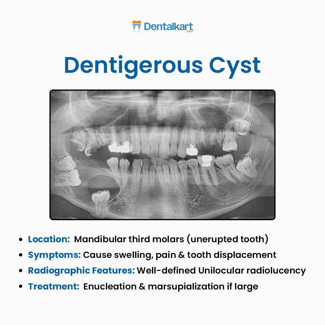
Dentigerous Cyst
Full Dental Analysis and Diagnosis
Location
Typically found around the mandibular third molars or maxillary canines.
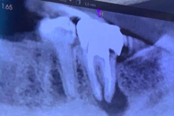
In this X-ray, the lesion appears surrounding the lower left third molar, expanding within the mandibular bone.
Radiographic Appearance
A well-defined unilocular radiolucency with a smooth, corticated border.
The cyst is attached to the cementoenamel junction (CEJ) of the unerupted tooth.
Some surrounding bone resorption is visible due to cystic expansion.
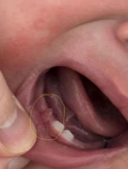
Symptoms and Clinical Signs
-
Swelling in the jaw region
-
Pain or pressure sensation as the cyst enlarges
-
Tooth displacement or root resorption of adjacent teeth
-
Sometimes asymptomatic and discovered incidentally on X-ray
Diagnosis
Provisional Diagnosis: Dentigerous Cyst associated with impacted third molar
Differential Diagnoses:
-
Odontogenic keratocyst (OKC)
-
Unicystic ameloblastoma
-
Radicular cyst (if adjacent to a non-vital tooth)
Confirmatory Test:
Histopathological examination after surgical removal confirms diagnosis.
Treatment Plan
1. Radiographic Evaluation
Perform panoramic and periapical radiographs to define cyst boundaries.
2. Surgical Intervention
-
Enucleation: Complete removal of the cystic lining and involved tooth (for moderate size cysts).
-
Marsupialization: For large cysts, to decompress gradually before full removal.
3. Follow-Up
Regular radiographs at 3, 6, and 12 months to ensure bone regeneration.
Maintain oral hygiene and prevent postoperative infection.
Healing Time Frame
-
Initial healing: 2–3 weeks (soft tissue)
-
Bone remodeling: 3–6 months
-
Full recovery: Up to 12 months depending on cyst size and bone loss
Issues That May Scale Up if Untreated
-
Expansion leading to jaw fracture
-
Secondary infection of the cyst cavity
-
Tooth root resorption of adjacent teeth
-
Possible transformation to ameloblastoma or other neoplastic lesions (rare)
-
Facial asymmetry or malocclusion
Comments
This case shows a classic dentigerous cyst presentation. It is essential to have it addressed promptly to prevent bone weakening and secondary infection. Early surgical management leads to excellent prognosis and bone regeneration.
Looking for dentist : Visit directory list
Teeth Case
 |
 |
 |
 |
 |
 |