Smile Again with Comfort and Confidence.
Dental Case Analysis: Unusual Bone-Like Structure Between Primary and Permanent Teeth in a Child
Image:
Severity:
Teeth Problems:
A Parent's Concern
Few things create more anxiety for parents than hearing a dentist say, "I'm not sure what this is."
When a child undergoes a routine dental examination and an X-ray reveals an unexpected structure inside the jaw, it can immediately raise concerns about tumors, abnormal bone growth, developmental disorders, or future problems with permanent teeth.
In this case, the dentist identified an unusual radiopaque (white) structure located between the developing permanent teeth and the primary teeth. Because the appearance was not typical, a referral to a specialist located approximately 150 miles away was recommended.
Naturally, the family wants to understand what they may be dealing with before making the long trip.
Important Disclaimer
A definitive diagnosis cannot be made from a single X-ray image alone.
A pediatric dentist, oral surgeon, or orthodontist will need to perform:
- Clinical examination
- Additional radiographs
- Possibly a CBCT scan (3D imaging)
- Review of the child's dental history
- Evaluation of tooth eruption patterns
However, we can discuss the most likely possibilities based on the radiographic appearance.
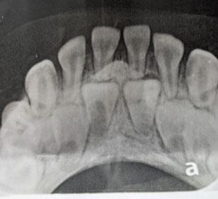
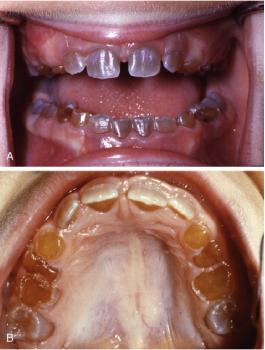
What Does the X-Ray Show?
The radiograph appears to show:
- Mixed dentition stage
- Developing permanent incisors
- Retained primary teeth
- An unusual radiopaque mass between the teeth
- Delayed eruption pathway of some permanent teeth
The structure appears denser than surrounding bone and is located in an area where normal tooth eruption should occur.
This is why the dentist likely recommended specialist evaluation.
Is This Really "Abnormal Bone Growth"?
Not necessarily.
Parents often describe these findings as "extra bone" because that is what they appear to be on X-rays.
However, several different conditions can create a similar appearance:
- Odontoma
- Supernumerary tooth
- Eruption obstruction
- Developmental dental anomaly
- Dense bone island
- Compound odontogenic lesion
Some are extremely common and benign.
Most Likely Possibility: Odontoma
One of the first conditions many specialists would consider is an odontoma.
What Is an Odontoma?
An odontoma is a benign developmental growth made from dental tissues.
It is not cancer.
It is not an infection.
It is not contagious.
It develops from the same tissues that form teeth.
Because of this, odontomas often contain:
- Enamel
- Dentin
- Cementum
- Pulp-like tissue
Essentially, the body creates extra tooth material in an abnormal shape.
Why Odontomas Are Often Found in Children
Odontomas frequently appear:
- During childhood
- During mixed dentition
- During permanent tooth eruption
Many children have no symptoms at all.
The condition is often discovered accidentally during routine dental X-rays.
Common Signs
Parents may notice:
- Permanent teeth not erupting
- Teeth coming in crooked
- Delayed tooth eruption
- Unusual spacing
- Retained baby teeth
The child may have absolutely no pain.
Types of Odontomas
Compound Odontoma
A compound odontoma contains many tiny tooth-like structures.
On X-rays they often resemble miniature teeth clustered together.
These commonly occur in the front of the upper jaw.
Complex Odontoma
A complex odontoma appears as a dense irregular mass.
Instead of tiny tooth shapes, it looks like a solid radiopaque structure.
This type can block normal tooth eruption.
Why Specialists May Suspect an Odontoma
Several features support this possibility:
- Child is in eruption age
- Structure appears radiopaque
- Location is near developing permanent teeth
- Eruption pathway may be obstructed
- Appearance differs from normal bone
This does not confirm the diagnosis, but it places odontoma high on the list of possibilities.
Another Possibility: Supernumerary Tooth
A supernumerary tooth is an extra tooth.
Some children develop more teeth than normal.
Mesiodens
The most common extra tooth is called a mesiodens.
It develops between the upper front teeth.
A mesiodens can:
- Block eruption
- Push teeth sideways
- Cause spacing issues
- Create crowding
Many are discovered during childhood.
How It Appears on X-Ray
A mesiodens often appears as:
- Tooth-shaped structure
- Cone-shaped tooth
- Inverted tooth
- Small malformed tooth
Sometimes it can be mistaken for other developmental anomalies.
Another Possibility: Eruption Obstruction
Permanent teeth must travel through bone before appearing in the mouth.
Anything blocking this pathway can create problems.
Examples include:
- Thick fibrous tissue
- Bone irregularities
- Odontomas
- Extra teeth
- Developmental defects
When eruption is delayed, dentists investigate for underlying causes.
Could It Be Cancer?
This is one of the biggest fears parents have.
Fortunately, based solely on this image, there are no obvious classic signs suggesting a malignant process.
Most childhood jaw cancers are extremely rare.
Features that often raise concern include:
- Rapid enlargement
- Bone destruction
- Severe pain
- Facial swelling
- Loose teeth
- Numbness
Those features are not apparent from this image alone.
However, specialist evaluation remains essential.
Could It Be an Infection?
The appearance does not strongly resemble a typical dental abscess.
Most abscesses appear as darker areas on radiographs due to bone loss.
This lesion appears relatively dense and radiopaque.
That makes infection less likely as the primary explanation.
Could It Be Normal Development?
Occasionally, developing permanent teeth overlap in unusual ways on 2D radiographs.
Because X-rays flatten three-dimensional structures into a two-dimensional image, anatomy can sometimes appear unusual.
A specialist may determine that what appears abnormal is actually overlapping structures.
Additional imaging helps clarify this.
Why a Specialist Referral Is Appropriate
Some parents worry when they hear their dentist say they don't know what something is.
In reality, this is often a sign of good professional judgment.
A dentist who recognizes an unusual finding and refers appropriately is acting responsibly.
Specialists have access to:
- Advanced imaging
- Pediatric dental expertise
- Oral pathology knowledge
- Orthodontic evaluation
- Surgical management if necessary
What Will the Specialist Likely Do?
Step 1: Review Medical History
Questions may include:
- Any genetic conditions?
- Delayed tooth eruption?
- Previous trauma?
- Family history of dental anomalies?
Step 2: Clinical Examination
The specialist will assess:
- Missing teeth
- Tooth mobility
- Swelling
- Gum condition
- Bite relationship
Step 3: Additional Imaging
Additional radiographs may include:
- Panoramic X-ray
- Occlusal radiograph
- CBCT scan
A CBCT scan provides a three-dimensional view.
This often reveals exactly what the structure is.
Step 4: Diagnosis
Once imaging is complete, the specialist can determine whether the finding represents:
- Odontoma
- Supernumerary tooth
- Developmental anomaly
- Dense bone island
- Other odontogenic lesion
Potential Treatment Options
Treatment depends entirely on the diagnosis.
Observation
If the lesion is harmless and not interfering with eruption:
- Periodic monitoring
- Follow-up X-rays
- No immediate treatment
may be recommended.
Surgical Removal
If an odontoma or extra tooth blocks eruption:
Minor oral surgery may be advised.
This is often performed under:
- Local anesthesia
- Sedation
- General anesthesia in younger children
Orthodontic Guidance
After removing the obstruction, orthodontic treatment may help guide permanent teeth into position.
Recovery After Surgical Removal
Most children recover quickly.
Typical recovery includes:
First 24 Hours
- Mild swelling
- Minor discomfort
- Soft diet
Days 2–7
- Healing progresses rapidly
- Swelling decreases
Weeks 2–4
- Soft tissue largely healed
Months Later
- Permanent teeth may begin erupting normally
What Happens If Left Untreated?
If the structure is blocking tooth eruption, delaying treatment could result in:
- Impacted permanent teeth
- Crowding
- Orthodontic complications
- Misalignment
- Delayed eruption
The longer the obstruction remains, the more difficult treatment may become.
Questions Parents Should Ask the Specialist
Before leaving the appointment, consider asking:
- What is the most likely diagnosis?
- Is this condition common?
- Does my child need a CBCT scan?
- Is surgery necessary?
- Will permanent teeth erupt normally?
- Could this affect future orthodontic treatment?
- What happens if we wait?
- How often should follow-up imaging be performed?
Reassurance for Parents
While the X-ray certainly shows something unusual, many conditions that create this appearance are benign and highly treatable.
In pediatric dentistry, the most common explanations are developmental dental anomalies such as:
- Odontomas
- Supernumerary teeth
- Eruption obstructions
These conditions are encountered regularly by pediatric dental specialists and oral surgeons.
The referral does not necessarily mean the condition is dangerous. More often, it means additional expertise and imaging are needed to determine exactly what is preventing normal tooth development.
Final Assessment
Based on the radiograph alone, the finding appears more consistent with a developmental dental anomaly than with infection or malignancy. The leading considerations include a compound odontoma, complex odontoma, or a supernumerary tooth (mesiodens-like structure) causing interference with the eruption of the permanent teeth.
Estimated Likelihood (Educational Opinion Only)
- Odontoma: High possibility
- Supernumerary tooth: Moderate to high possibility
- Eruption-related developmental anomaly: Moderate possibility
- Dense bone island: Lower possibility
- Infection: Lower possibility
- Malignancy: Appears unlikely based on this image alone
A pediatric dental specialist should evaluate the child as soon as practical, but this X-ray does not immediately suggest a dental emergency. Most similar cases are successfully treated with monitoring or minor surgical intervention, allowing the permanent teeth to continue developing normally.
"Our 5-Year-Old Child Has Some Issues Our Dentist Has Never Seen Before"
After reviewing the Reddit discussion, the overwhelming consensus from multiple dentists, including a pediatric dentist, was that the X-ray likely does not show abnormal bone growth at all. Most commenters believed the image quality and angle of the X-ray created the appearance of an unusual structure.
Main Opinion From Most Dentists
The majority of dental professionals stated:
- The X-ray is poorly angled.
- The white area appears to be normal chin bone anatomy overlapping the image.
- There is no obvious evidence of a tumor, cyst, extra bone growth, or serious pathology.
- The appearance is likely an X-ray artifact caused by positioning.
Several comments were very direct:
"Nothing wrong here."
"This is just a terrible x-ray."
"That's just part of the chin."
Pediatric Dentist's Opinion
A verified pediatric dentist joined the discussion and stated that the structure appears to be:
- Normal anatomy
- Part of the chin
- Visible because the X-ray was taken at an incorrect angle
The pediatric dentist agreed with the other professionals that there was likely nothing unusual present.
Why It Looks Strange
According to several dentists:
The X-ray appears to be an occlusal film taken at an excessive angle.
This causes:
- Chin bone superimposition
- Overlapping structures
- Distorted appearance
As a result, normal anatomy can appear as a dense mass between developing permanent teeth.
Minority Opinion: Still Keep the Referral Appointment
Although most dentists believed the image was normal, one experienced commenter recommended still attending the specialist appointment.
Their concern was not the "bone growth" itself.
Instead, they noted:
- Possible differences in the pulp chambers of the permanent central incisors
- Slight irregularities around the developing teeth
- Difficulty making a complete diagnosis from a single low-quality X-ray
They suggested additional evaluation might still be worthwhile.
Another Conservative View
One commenter noticed that the structure appeared to be in the eruption path of the permanent incisors.
Their recommendation:
- Monitor the child
- Take another X-ray in 6–12 months
- Verify that permanent teeth continue erupting normally
They did not believe it was an emergency.
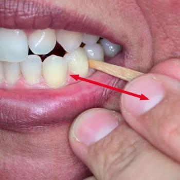
Parent Follow-Up Information
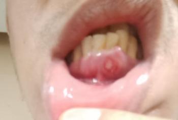
The parent later commented that:
"It does look ok but there is a nodule you can feel just below the gum line."
This caused some additional discussion.
However, even after learning about the palpable bump, most professionals still believed the radiographic appearance represented normal anatomy or projection error rather than disease.
Overall Community Consensus
Most Likely Explanation
- Normal chin bone anatomy
- X-ray overlap
- Imaging artifact
- Poor X-ray angle
Less Likely
- Odontoma
- Supernumerary tooth
- Abnormal bone growth
Very Unlikely Based on Comments
- Cancer
- Aggressive lesion
- Serious jaw pathology
Recommended Action
- Keep the specialist appointment if feasible.
- Obtain better quality imaging.
- Consider a repeat occlusal X-ray or CBCT if the specialist feels it is necessary.
- Monitor eruption of the permanent incisors.
Teeth Case
 |
 |
 |
 |
 |
 |