Healthy Teeth, Happy Life.

Dentinogenesis Imperfecta: Management of Severe Primary Tooth Abrasion and Enamel Fracture in Mixed Dentition
Image:
Severity:
Teeth Problems:
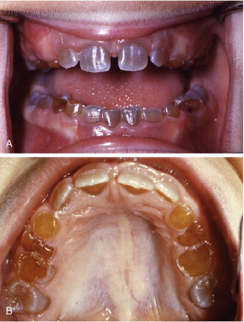
Zoom 100% & Deep Examination (Detailed Color Analysis)
| Feature | Observation (Image A & B) | Analysis based on Dentinogenesis Imperfecta (DI) |
| Primary Teeth (Deciduous) | Severely abraded, flat, and worn down (Image B, showing upper primary molars). Color appears amber/yellow-brown (opalescent) in Image B. | This severe wear is characteristic of DI. The poorly mineralized dentin and weakened dentin-enamel junction cause the brittle enamel to fracture easily, leading to rapid wear of the underlying dentin. |
| Lower Permanent Incisors | The biting (incisal) edges are very translucent/opalescent (Image A), and the enamel appears to be chipping or breaking away. | This is the defining feature of DI: "Hereditary Opalescent Dentin." The abnormal dentin is responsible for the gray/bluish-brown or amber color and translucency. The enamel loss is due to the lack of proper support from the abnormal dentin. |
| Overall Color | A mix of white/translucent (permanent teeth enamel breaking away) and deep amber/brown/yellow (exposed, abnormal primary dentin). | The different colors reflect the different stages of the disease—the abnormal dentin is what gives DI its characteristic opalescent color. |
Full Analysis and Diagnosis
-
Condition: Dentinogenesis Imperfecta (Type I or II)—A hereditary developmental disorder of the dentin.
-
Pathology: The dentin structure is defective, resulting in teeth that are often discolored, bulbous-shaped, and structurally weak.
-
Clinical Problems:
-
Enamel Fracture: The abnormal dentin-enamel junction leads to brittle enamel that fractures easily, exposing the soft, poorly mineralized dentin.
-
Severe Abrasion: The exposed dentin wears down rapidly, leading to loss of vertical dimension, affecting chewing function, and potentially damaging the permanent teeth as they erupt.
-
Pulp Exposure/Necrosis: The severe wear can quickly lead to pulp exposure and infection, even if the pulp chambers are often initially obliterated (a common finding in DI).
-
Clinical Management of Severe Enamel Fracture in Children With Dentinogenesis Imperfecta
Why Early Dental Protection Matters
Severe tooth wear and repeated enamel fractures in a child with dentinogenesis imperfecta (DI) require more than routine restorative treatment. This inherited condition affects dentin formation, making the tooth structure less able to support the overlying enamel. As the enamel fractures or separates, the exposed dentin may wear down rapidly under normal chewing forces.
In the primary dentition, extensive tooth destruction can develop before parents recognize the underlying condition. When permanent teeth begin erupting, the dentist must also consider long-term tooth preservation, the child's growth, and the development of a stable bite.
The clinical priority is to preserve healthy tooth structure, protect the dental pulp, maintain chewing function, and prevent avoidable tooth loss. Treatment should be individualized according to the severity of structural damage, the child's age, symptoms, and stage of dental development. Complex cases may benefit from referral to a pediatric dentist with experience managing inherited dental disorders.
1. Distinguishing Dentinogenesis Imperfecta From Isolated Enamel Fracture
An enamel fracture does not automatically mean a child has DI. Trauma, developmental enamel defects, tooth decay, and other dental conditions can produce similar findings.
The clinical distinction is important:
-
Dentinogenesis imperfecta: Abnormal dentin development can compromise the support beneath the enamel, allowing enamel to fracture and dentin to wear down.
-
Amelogenesis imperfecta: The primary abnormality affects enamel formation, thickness, or mineralization.
-
Traumatic enamel fracture: A tooth may chip following an impact or biting injury without an underlying inherited disorder.
-
Dental caries: Bacterial acid activity damages tooth structure and may weaken the remaining enamel and dentin.
Teeth affected by DI may appear blue-gray, amber, or brown, although appearance varies. Some patients have substantial wear, while others have relatively mild visible changes. A family history of similar dental problems can provide an important diagnostic clue.
The dentist should evaluate the entire dentition rather than focusing only on the most visibly damaged tooth.
2. Diagnostic Assessment of Severe Primary-Tooth Wear
A structured assessment helps determine whether the tooth can be restored and whether the dental pulp remains healthy.
Clinical examination
The examination should document:
-
The location and extent of enamel fractures.
-
The amount of exposed dentin and remaining tooth structure.
-
Tooth discoloration and unusual crown shape.
-
Sensitivity to temperature, touch, or chewing.
-
Signs of pulp exposure, inflammation, infection, or abscess.
-
The child's bite, chewing efficiency, and ability to eat comfortably.
-
The eruption pattern of permanent teeth.
The dentist should also ask when the fractures began, whether they followed an injury, and whether other family members have similar dental findings.
Dental imaging
Radiographs may help evaluate root development, pulp chamber and root canal appearance, periapical tissues, and the condition of developing permanent teeth. DI can be associated with characteristic changes in the shape of the pulp chambers and roots, but findings vary between individuals.
Radiographs should be selected according to the child's clinical needs. They should not be taken automatically at every visit or used as a substitute for a clinical examination.
Assessing the dental pulp
A tooth with extensive structural loss is not necessarily infected or non-vital. Conversely, a tooth that appears relatively intact externally may have significant internal pathology.
The dentist must distinguish among a restorable tooth with healthy or reversibly inflamed pulp, a tooth requiring vital pulp therapy, and a tooth with irreversible pulp disease or necrosis. This distinction determines whether protective restoration, pulp treatment, or extraction is appropriate.
3. Treatment Options for Severe Primary-Tooth Abrasion and Fracture
Treatment should be selected according to the remaining tooth structure, pulp condition, expected time before natural exfoliation, and the child's ability to tolerate the procedure.
Fluoride and sensitivity management
Professional fluoride varnish and suitable desensitizing measures may help reduce sensitivity and support preventive care. Fluoride does not rebuild missing enamel or correct the underlying genetic dentin defect, so severe structural damage usually requires additional treatment.
Tooth-colored restorations
Composite resin or other suitable restorative materials may be considered for localized defects when adequate tooth structure remains to support the restoration.
The dentist must consider moisture control, the bonding surface, the forces placed on the tooth, and the likelihood of further fracture. Restorations may require repair or replacement over time.
Full-coverage crowns for primary teeth
When a primary molar has extensive wear or weakened walls, a preformed stainless steel crown may offer more comprehensive protection than a small filling. The decision depends on the tooth's restorability, pulp status, remaining root structure, and expected service life.
For severely damaged primary anterior teeth, tooth-colored restorative options or selected pediatric crowns may be considered when clinically appropriate. Aesthetic treatment must not compromise pulp health or unnecessarily remove remaining tooth structure.
Pulp therapy or extraction
If extensive fracture or wear has affected the pulp, treatment may involve a vital pulp procedure or other pulp therapy, depending on the diagnosis and restorability of the tooth.
Extraction may be necessary when a tooth cannot be predictably restored, has an unfavorable prognosis, or presents an infection that cannot be appropriately managed by preserving it. The dentist should also consider the child's age, the tooth's role in maintaining space, and the possible effects on the developing dentition.
No single treatment is appropriate for every child with DI. The American Academy of Pediatric Dentistry provides guidance on pediatric restorative treatment and pulp therapy that can inform these decisions.
4. Protecting Newly Erupted Permanent Teeth
Mixed dentition—the period when primary and permanent teeth coexist—requires a preventive strategy as well as treatment of existing damage.
Newly erupted permanent teeth may still have incomplete root development. Preserving pulp vitality is particularly important because a healthy pulp supports continued root formation.
Recommended management may include:
-
Monitoring the eruption and structural integrity of permanent teeth.
-
Providing fluoride-based prevention and individualized oral hygiene instruction.
-
Considering sealants for suitable vulnerable tooth surfaces.
-
Restoring fractures early when protection or function is compromised.
-
Using partial-coverage or full-coverage restorations only when indicated by the extent of damage and the tooth's developmental stage.
-
Reviewing the bite and distribution of chewing forces.
-
Coordinating long-term care with a pediatric dentist and other specialists when necessary.
Extensive definitive restorative work may need to be staged as the child grows. Permanent crowns and other indirect restorations should be planned with attention to tooth development, gum health, future treatment needs, and the expected lifespan of each restoration.
5. Home Care and Prevention of Further Tooth Damage
Home care cannot reverse DI, but it can reduce additional damage from preventable causes and help protect vulnerable teeth.
Practical recommendations for parents:
-
Brush twice daily with an age-appropriate amount of fluoride toothpaste.
-
Supervise brushing when the child needs help cleaning effectively.
-
Limit frequent sugary snacks and drinks.
-
Reduce frequent exposure to acidic drinks, which can contribute to additional tooth wear.
-
Avoid chewing ice, very hard sweets, and other objects that may fracture weakened teeth.
-
Cut firm foods into manageable pieces when biting causes discomfort.
-
Maintain balanced nutrition with adequate calcium and vitamin D intake as part of a healthy diet.
-
Keep regular dental appointments even when the child has no pain.
Children with severe DI may need professional reviews every three to six months, or more frequently when wear is progressing, restorations are failing, or symptoms are present. The appropriate interval should be determined by the treating dentist.
6. When Is Severe Enamel Fracture an Urgent Dental Problem?
A newly fractured tooth should be assessed promptly, especially when dentin is exposed or the child reports pain.
Arrange urgent dental evaluation for:
-
A large fracture exposing deep tooth structure.
-
Persistent or spontaneous toothache.
-
Pain that continues after hot or cold stimulation.
-
Pain during chewing that prevents normal eating.
-
A visible pink or red area within a fracture, which may indicate pulp exposure.
Facial or gum swelling, pus, fever associated with dental symptoms, or rapidly worsening pain requires urgent assessment. Difficulty swallowing or breathing, or swelling that is spreading rapidly toward the eye or neck, requires emergency medical care.
Antibiotics alone do not repair a fractured tooth or eliminate the source of a dental infection. When infection is present, definitive treatment may require drainage, pulp treatment, or extraction, depending on the diagnosis.
7. Long-Term Prognosis
Dentinogenesis imperfecta is a lifelong structural condition, but it does not mean that all affected teeth will inevitably be lost.
The prognosis depends on the severity of the defect, the timing of diagnosis, pulp health, the amount of remaining tooth structure, the success of protective restorations, and continued follow-up.
A successful treatment plan should evolve with the child. The immediate objective is to relieve symptoms and protect vulnerable teeth. The longer-term objective is to preserve function, maintain a healthy bite, protect permanent teeth, and plan appropriate restorative care through adolescence and adulthood.
Some forms of DI occur as part of osteogenesis imperfecta, while others are predominantly dental. If the medical history or clinical findings suggest a broader inherited condition, medical or genetic assessment may be appropriate.
Frequently Asked Questions: Dentinogenesis Imperfecta and Severe Tooth Fracture
1. Can dentinogenesis imperfecta cause severe enamel fractures in baby teeth?
Yes. Abnormal dentin may provide inadequate support for the enamel. As the enamel fractures, the exposed dentin can wear down rapidly during ordinary chewing. The degree of damage varies from child to child.
2. Is severe tooth abrasion the same as enamel erosion?
No. Abrasion generally refers to mechanical wear caused by external forces, while erosion is chemical loss of tooth structure caused by acids. Attrition is wear caused by tooth-to-tooth contact. Children with DI may experience several types of tooth wear simultaneously, so the dentist should identify contributing factors before selecting treatment.
3. Can a dentist save a severely worn primary tooth?
Often, yes, provided the tooth has enough remaining structure and a treatable pulp condition. Depending on the case, treatment may involve a restoration, a protective crown, or pulp therapy followed by restoration. Extraction is considered when the tooth cannot be predictably preserved.
4. Do all children with DI need dental crowns?
No. Crowns are not automatically required for every affected tooth. They may be appropriate when extensive wear or fracture makes a small restoration unreliable, particularly in primary molars. Less extensive defects may be managed with more conservative treatment.
5. Can fluoride repair broken enamel?
No. Fluoride can help prevent decay and support remineralization of early mineral loss, but it cannot regenerate a missing portion of enamel or correct defective dentin formation. Structural fractures may require restorative protection.
6. Will permanent teeth also be affected?
They may be. DI can affect both primary and permanent teeth, although the severity varies. Newly erupted permanent teeth should be examined and monitored so that protective treatment can be provided when necessary.
7. Does a fractured tooth always need a root canal?
No. A fracture does not automatically mean the pulp is irreversibly damaged. The dentist must assess the depth of the fracture, pulp symptoms, clinical findings, and appropriate radiographs. When possible, preserving a healthy pulp is especially important in immature permanent teeth.
8. Should a child with DI avoid all hard foods?
Not necessarily. A restrictive diet is rarely the goal. Parents should avoid particularly hard foods or biting habits that trigger fractures and adapt food texture when necessary. The child should still receive balanced nutrition, with dietary advice individualized to the severity of tooth damage.
9. How often should a child with severe DI visit the dentist?
Children with active tooth wear, extensive restorations, or recurring fractures may need reviews approximately every three to six months. The dentist may recommend shorter intervals if the condition is progressing or symptoms develop.
10. Can dentinogenesis imperfecta be cured?
There is currently no treatment that corrects the inherited dentin defect throughout the teeth. However, preventive care, timely restorations, and long-term monitoring can help maintain comfort, appearance, and chewing function.
11. Is DI always associated with osteogenesis imperfecta?
No. Some forms of DI are associated with osteogenesis imperfecta, a condition affecting bones and connective tissue, while other forms primarily affect the teeth. A medical history and appropriate assessment help determine whether additional evaluation is needed.
12. When should parents seek emergency care for a fractured tooth?
Seek prompt dental care for significant fractures, severe pain, or suspected pulp exposure. Facial swelling, fever with dental infection, or rapidly worsening symptoms need urgent evaluation. Difficulty breathing or swallowing, or rapidly spreading facial or neck swelling, requires emergency medical care.
Clinical Takeaway
Severe primary-tooth abrasion and enamel fracture in a child with suspected dentinogenesis imperfecta should trigger a comprehensive assessment rather than repeated patch repairs alone. The most appropriate strategy combines early diagnosis, pulp assessment, tooth-appropriate restorative protection, prevention of additional wear, and long-term monitoring of permanent tooth development.
Treatment decisions should be made by the child's treating dentist based on the clinical examination, appropriate imaging, and individual needs. These recommendations are educational and do not replace an in-person dental diagnosis.
Clinical References
-
American Academy of Pediatric Dentistry. Pediatric Restorative Dentistry. AAPD clinical guidance.
-
American Academy of Pediatric Dentistry. Pulp Therapy for Primary and Immature Permanent Teeth: Indications and Objectives. AAPD clinical guidance.
-
American Academy of Pediatric Dentistry. Management of the Developing Dentition and Occlusion in Pediatric Dentistry. AAPD clinical guidance.
Process to Execute (Treatment Plan)
The goal is to protect the teeth from wear, maintain function, and preserve vertical dimension. This is a long-term, multi-stage process:
-
Immediate Protection of Primary Teeth:
-
Full-coverage restorations (e.g., Stainless Steel Crowns (SSCs)) for the primary molars to prevent further abrasion and maintain the bite space until the permanent teeth erupt.
-
Composite or resin restorations may be used on anterior teeth to protect the exposed dentin and prevent sensitivity.
-
-
Protection of Permanent Teeth (As they Erupt):
-
Prompt placement of full-coverage restorations (crowns), often ceramic or composite, on permanent teeth (especially incisors and molars) immediately after they erupt and the gum tissue has matured. This prevents the enamel from fracturing and the underlying dentin from wearing away.
-
-
Long-Term Management:
-
Restoration of Vertical Dimension: As the child grows, a final full-mouth rehabilitation (using crowns, fixed bridges, or possibly implants later in adulthood) will be necessary to restore the height and function of the bite.
-
Orthodontic Consultation: May be required to align the bite properly for the final restorations.
-
Time Frame to Heal & If Takes 14 Days
-
No Healing Time: DI is a genetic, non-curable condition. The "healing" concept is replaced by "management" and "protection."
-
Time Frame for Treatment: The management is lifelong.
-
Placing protective crowns/restorations on primary teeth may take 2-4 dental visits.
-
Protecting the permanent teeth is an ongoing process over many years as they erupt.
-
What Will Be the Issue That Will Scale Up (If rushed or ignored)?
If the teeth are not protected quickly, the issue will scale up rapidly:
-
Accelerated Tooth Loss: Unprotected, the teeth will wear down extremely fast, leading to total loss of tooth structure and bite height (vertical dimension).
-
TMJ Problems: The loss of vertical dimension forces the jaw joint (TMJ) into an unnatural position, causing pain, clicking, and long-term joint damage.
-
Pulp Necrosis and Infection: The wear will quickly expose the pulp (nerve) of the teeth, leading to infection, abscesses, and the need for root canals or extractions.
-
Psychosocial Impact: The severe discoloration and poor appearance can significantly affect the child's self-esteem and social development.
Comments
This case highlights the critical need for early diagnosis and aggressive, preventive restoration (e.g., full-coverage crowns) to manage Dentinogenesis Imperfecta. The protective restorations act as a physical shield against rapid wear, which is the most destructive aspect of the condition.
Visit Nearest Location Area
Given the complexity of this genetic condition, it is vital to see a specialist, ideally a Pediatric Dentist or a Prosthodontist experienced in managing DI.
You can use the directory listing to start your search:
https://cebudentalimplants.com/map-dental-clinic
Expert Dental Guidance: Managing Dentinogenesis Imperfecta and Severe Tooth Wear
Dentinogenesis imperfecta (DI) is a hereditary disorder affecting the formation and structure of dentin, the hard tissue beneath tooth enamel. Teeth affected by DI may appear unusually translucent, blue-gray, yellow-brown, or amber and can be more susceptible to enamel fracture, rapid tooth wear, and sensitivity.
Because the severity varies considerably between individuals, treatment should be individualized after a comprehensive examination by a dentist. In children, early evaluation is particularly important because protecting the remaining tooth structure can help maintain comfort, chewing function, appearance, and normal oral development.
What dentists look for
A dentist may evaluate:
-
Tooth color and translucency
-
Enamel chipping or fracture
-
Abnormal tooth wear or loss of tooth structure
-
Dentin exposure and sensitivity
-
Dental abscesses or signs of infection
-
Existing restorations and their condition
-
The child's ability to chew comfortably
-
Oral hygiene and plaque accumulation
-
The development and eruption of permanent teeth
-
Family history of similar dental findings
Dental X-rays may also be recommended. Depending on the type and severity of DI, radiographs can show characteristic changes in the shape and structure of the teeth.
Why early dental care matters
Severely affected teeth can lose protective enamel and become increasingly vulnerable to wear and fracture. However, severe tooth wear does not automatically mean that a tooth must be extracted.
A dentist should determine whether the tooth can be maintained using preventive care, protective restorations, or other conservative treatment. Extraction may sometimes be necessary when a tooth cannot be predictably restored, has extensive infection, or is causing other significant problems.
For children, treatment planning should also consider growth, eruption of permanent teeth, space management, and long-term oral development.
Dentist Expert Perspective
Clinical principle: The goal is not simply to repair damaged teeth. The dentist should try to preserve healthy tooth structure, control symptoms, prevent further breakdown, and maintain oral function while considering the patient's age and future dental development.
A dentist may recommend a combination of preventive and restorative measures depending on the individual case. These can include fluoride-based preventive care, careful oral hygiene, dietary guidance, protective restorations, treatment of sensitivity, and regular monitoring.
For children with substantial tooth destruction, a pediatric dentist may be particularly helpful because treatment must account for primary teeth, permanent tooth development, behavior management, growth, and the timing of natural tooth exfoliation.
Important medical caution
There is no single treatment protocol that is appropriate for every person with dentinogenesis imperfecta. Treatment depends on the patient's age, the teeth affected, the amount of remaining tooth structure, symptoms, infection, bite, oral hygiene, and the condition of the permanent dentition.
Online photographs cannot reliably determine whether a tooth can be saved. A clinical examination and appropriate dental imaging are needed before making treatment decisions.
Frequently Asked Questions (FAQ): Dentinogenesis Imperfecta, Severe Primary Tooth Abrasion, and Enamel Fracture
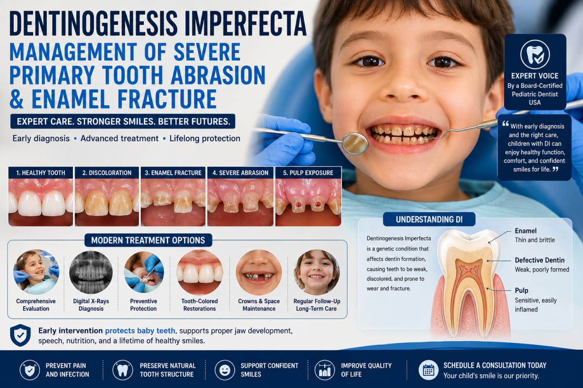
Expert Answers by a Board-Certified Pediatric Dentist (USA)
1. What is Dentinogenesis Imperfecta (DI)?
Expert Answer:
Dentinogenesis Imperfecta (DI) is a rare inherited dental condition that affects how dentin—the layer beneath the enamel—develops. Although the enamel may appear normal when a tooth first erupts, it lacks proper support because the underlying dentin is weak and poorly formed.
As a result, teeth become fragile and are prone to:
- Rapid enamel fracture
- Severe tooth wear (abrasion and attrition)
- Tooth discoloration ranging from blue-gray to amber-brown
- Increased sensitivity
- Early loss of tooth structure
In my pediatric practice, early diagnosis is essential because children with DI often experience significant tooth wear long before their permanent teeth fully erupt.
2. What are the first signs parents should watch for?
Expert Answer:
Many parents initially think their child's teeth are simply stained or naturally dark.
Common early signs include:
- Blue-gray, yellow-brown, or amber-colored teeth
- Front teeth that chip easily
- Flat chewing surfaces
- Rapid tooth wear despite good brushing
- Teeth appearing shorter over time
- Small fractures after biting soft foods
- Increased sensitivity to cold foods or drinks
Children with these symptoms should receive a comprehensive dental evaluation as soon as possible.
3. Why do the teeth wear down so quickly?
Expert Answer:
Unlike healthy teeth, those affected by Dentinogenesis Imperfecta have defective dentin that cannot adequately support the enamel.
Once the enamel chips away, the softer dentin is exposed and wears down rapidly during everyday chewing.
Because baby teeth naturally have thinner enamel than permanent teeth, this process may occur much faster in young children.
Without treatment, entire biting surfaces may disappear within a relatively short period.
4. Can Dentinogenesis Imperfecta affect both baby teeth and permanent teeth?
Expert Answer:
Yes.
DI commonly affects both the primary (baby) and permanent dentitions.
In many children, the baby teeth show the earliest and most severe changes, allowing dentists to diagnose the condition before permanent teeth erupt.
Early diagnosis gives us an opportunity to protect the developing permanent teeth with preventive care and long-term treatment planning.
5. Is Dentinogenesis Imperfecta inherited?
Expert Answer:
Yes.
Most cases are inherited in an autosomal dominant pattern, meaning a child can inherit the condition if one parent carries the altered gene.
Because of its genetic nature, dentists often recommend evaluating siblings and reviewing the family's dental history.
Genetic counseling may also be appropriate for affected families.
6. What happens if this condition is left untreated?
Expert Answer:
Without treatment, Dentinogenesis Imperfecta can lead to serious oral health problems.
Potential complications include:
- Progressive enamel fractures
- Severe tooth abrasion
- Pulp exposure
- Tooth sensitivity progressing to pain
- Dental infections
- Gum inflammation
- Difficulty chewing
- Poor nutrition
- Speech development challenges
- Premature loss of primary teeth
- Bite abnormalities
- Emotional distress due to the appearance of the teeth
Early intervention greatly reduces these risks.
7. How do dentists diagnose Dentinogenesis Imperfecta?
Expert Answer:
Diagnosis combines several clinical and radiographic findings.
A pediatric dentist typically performs:
- Comprehensive oral examination
- Family medical and dental history review
- Digital dental X-rays
- Bite evaluation
- Assessment of tooth color and wear patterns
- Evaluation for enamel fractures
- Monitoring eruption of permanent teeth
Radiographs often reveal characteristic changes such as bulbous crowns, shortened roots, and partial or complete pulp chamber obliteration.
8. What treatment options are available?
Expert Answer:
Treatment depends on the child's age, severity of tooth wear, and stage of dental development.
Management may include:
- Fluoride varnish applications
- Desensitizing agents
- Tooth-colored composite restorations
- Glass ionomer restorations
- Stainless steel crowns for primary molars
- Pediatric zirconia crowns for front teeth in selected cases
- Protective bite appliances when indicated
- Regular professional monitoring
- Long-term restorative planning as permanent teeth erupt
The primary goal is preserving tooth structure while maintaining comfort, function, and appearance.
9. Will my child eventually need crowns?
Expert Answer:
Many children with moderate to severe DI benefit from full-coverage restorations.
Crowns protect weakened teeth by:
- Preventing additional fractures
- Restoring chewing function
- Reducing sensitivity
- Improving appearance
- Preserving remaining tooth structure
The type of crown depends on which teeth are affected and the child's age.
10. Can children with Dentinogenesis Imperfecta eat normally?
Expert Answer:
Yes, but dietary adjustments often help protect weakened teeth.
We generally recommend:
- Softer nutritious foods
- Limiting very hard candies
- Avoiding chewing ice
- Avoiding biting directly into hard fruits
- Cutting foods into smaller pieces
- Drinking water instead of acidic beverages
Proper nutrition remains extremely important because growing children require balanced diets for healthy development.
11. How often should children with DI visit the dentist?
Expert Answer:
Children with Dentinogenesis Imperfecta usually require closer monitoring than children with healthy teeth.
Many pediatric dentists recommend examinations every 3 to 6 months, depending on disease severity.
Frequent visits allow us to:
- Monitor tooth wear
- Repair small fractures early
- Apply fluoride protection
- Adjust restorations
- Evaluate eruption of permanent teeth
- Prevent major complications before they occur
12. Can permanent teeth be protected before they become damaged?
Expert Answer:
Absolutely.
One of the biggest advantages of early diagnosis is preventive planning.
As permanent teeth erupt, dentists can implement protective measures such as:
- Regular fluoride therapy
- Dental sealants when appropriate
- Early restorative care
- Customized oral hygiene instruction
- Bite monitoring
- Protective appliances for children with excessive tooth grinding
These strategies significantly improve the long-term prognosis.
13. Is Dentinogenesis Imperfecta painful?
Expert Answer:
Not always during the earliest stages.
However, as enamel fractures expose dentin and wear progresses, many children develop sensitivity to cold foods, sweet drinks, and chewing pressure.
If decay or fractures reach the dental pulp, pain may become severe and may require more complex treatment.
Early management often prevents discomfort before it develops.
14. What is the long-term outlook for children with Dentinogenesis Imperfecta?
Expert Answer:
With early diagnosis, individualized treatment, and regular follow-up, the outlook is very encouraging.
Modern pediatric dentistry focuses on preserving natural tooth structure, protecting developing permanent teeth, restoring normal chewing and speech, and improving aesthetics throughout childhood. While Dentinogenesis Imperfecta cannot be cured because it is a genetic condition, timely preventive care and restorative treatment allow most children to maintain healthy function, avoid many complications, and enjoy confident smiles as they grow into adulthood.
Related Clinical Cases
Patients with lower front gum inflammation may also experience tartar buildup, periodontal disease, gum abscesses, or tooth decay. Explore these related clinical case studies.



Other topics:
Other Dental Clinic
 |
 |
 |
 |