How to Prevent Dry Socket After Tooth Extraction (Expert Guide 2025)
Topics:
Dry socket is one of the most common complications after a tooth extraction—and also one of the most preventable. It occurs when the protective blood clot in the socket is dislodged or fails to form, exposing the bone and nerves underneath.
Dental organizations and clinical experts emphasize that proper aftercare in the first 3–5 days is critical to prevent this painful condition.
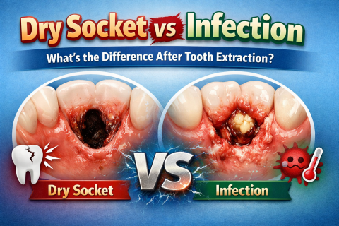
What Is Dry Socket and Why It Happens
Dry Socket (also called alveolar osteitis) develops when the healing process is interrupted.