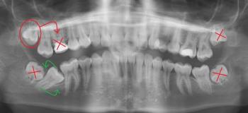
Post-root canal pain

Severe Night Toothache & Deep Cavity Treatment in California, US

Severity: 

The image strongly suggests a case of Severe Tooth Decay and Possible Pulp Exposure. The pain shown in the right panel indicates a severe toothache, which often means the decay has progressed to the innermost part of the tooth (the pulp), causing irreversible pulpitis or a dental abscess. This requires urgent professional attention.

Do not attempt to self-treat this condition. Use the provided directory link to find a nearby clinic immediately.


Lower anterior cervical roughness - likely calculus or non-carious wear; cleaning and possible restoration recommended

Severity: 

Quick clinical impression

  • The lower anterior teeth show rough, yellowish/cream areas at the cervical (gumline) region of several incisors.

  • Most likely causes (photo only — no probing/X-ray): tartar/calculus buildup with superficial stain, and/or non-carious cervical wear (abrasion/erosion) exposing dentine. The rough texture could be hardened calculus or eroded tooth surface.

Deep Interproximal Cavities & Restorations: Immediate Treatment Plan + 14-Day Recovery Guide

Full analysis & diagnosis (Zoom 100%)

  • Findings from the images: Rubber dam isolation with matrix bands in place; multiple deep occlusal-/proximal caries excavations on posterior teeth. Some restorations are in progress — there is close proximity to the pulp on at least one surface. Tooth structure shows large cavities with thin remaining walls.

How Fast Caries Can Progress - From Filling to Root Canal in 5 Months

Severity: 

Nice clear radiographs — thanks. I’ll give a focused clinical/radiographic read, a 0–14 day action plan you can use now, likely definitive options, what will get worse if ignored, and short practical comments for the patient and clinician.


Radiographic diagnosis (concise)

  • The images show a deep carious lesion extending into the pulpal chamber of a posterior tooth (likely a mandibular molar from the root morphology).

Pages