Strong Teeth, Fresh Breath, Brighter You.

Why Lower Molars Fail More Often: A USA Dental Expert Explains the Science, Clinical Risks, and How to Save Your Back Teeth
Language :
Topics:
Among all permanent teeth, lower molars (mandibular molars) perform the majority of chewing, grinding, and crushing food. Every day, they withstand tremendous biting pressure while simultaneously being exposed to bacteria, food debris, saliva, and acidic by-products.
Unfortunately, these same characteristics make lower molars one of the most commonly damaged teeth in clinical dentistry.
Across dental practices in the United States, dentists frequently see patients requiring fillings, root canal therapy, crowns, periodontal treatment, or even extraction because disease has progressed silently within these back teeth. By the time pain develops, extensive damage has often already occurred.
Understanding why lower molars fail more often helps patients recognize early warning signs and seek treatment before minor problems become complex and expensive.
USA Clinical Case Review: Severe Lower Molar Decay with Periodontal Involvement
A 42-year-old patient presented to a U.S. dental clinic complaining of:
- Severe localized pain while chewing
- Sensitivity to cold beverages
- Food becoming trapped around the back lower tooth
- Occasional gum bleeding during brushing
- Increasing discomfort over several weeks
Clinical Examination
A comprehensive oral examination and digital radiographs revealed:
- Extensive occlusal-proximal dental decay
- Significant enamel destruction
- Deep dentin involvement approaching the pulp
- Localized gingival inflammation
- Bleeding on periodontal probing
- Deep periodontal pocket surrounding the affected molar
- Early supporting bone loss adjacent to the roots
Fortunately, the tooth remained structurally restorable because treatment was initiated before catastrophic root fracture occurred.
Following removal of infected tooth structure, periodontal therapy, and definitive restoration, the patient's pain resolved and chewing function was successfully restored.
This case illustrates why lower molars require immediate evaluation when symptoms first appear. Delayed treatment greatly increases the risk of root canal therapy, surgical procedures, or tooth loss.
Why Are Lower Molars More Vulnerable?
Unlike front teeth, mandibular molars possess unique anatomical and functional characteristics that significantly increase their susceptibility to disease.
These include:
- Complex chewing surfaces
- Heavy biting forces
- Difficult cleaning access
- Multi-root anatomy
- Increased plaque accumulation
- Food impaction
- Greater periodontal complexity
Together, these factors create an ideal environment for bacterial colonization and progressive dental disease.
1. Deep Pits and Fissures Trap Bacteria
The chewing surface of every lower molar contains intricate grooves called pits and fissures.
These microscopic depressions are designed to grind food efficiently, but they also trap:
- Food particles
- Sugars
- Bacteria
- Plaque biofilm
Many fissures are actually narrower than toothbrush bristles, preventing effective cleaning during routine brushing.
As bacteria metabolize sugars, they produce acids that gradually dissolve enamel through a process known as demineralization.
Decay frequently begins deep within these grooves before becoming visible externally.
2. Lower Molars Absorb the Greatest Chewing Forces
Lower molars sit closest to the jaw joint, allowing them to generate extraordinary biting power.
Studies demonstrate that posterior teeth experience the highest occlusal loads during chewing.
These forces repeatedly stress:
- Tooth enamel
- Existing fillings
- Dental crowns
- Internal dentin
- Root structures
If decay weakens the tooth, normal chewing can cause:
- Cracked cusps
- Fractured restorations
- Split teeth
- Vertical root fractures
Even a small untreated cavity may rapidly progress into catastrophic structural failure because of these constant mechanical stresses.
3. They Are Difficult to Clean Properly
Lower molars are positioned at the very back of the mouth.
Several anatomical factors make effective cleaning challenging:
- Limited mouth opening
- Tongue interference
- Buccinator muscle pressure
- Reduced visibility
- Limited toothbrush angle
As a result, plaque commonly remains on:
- Distal surfaces
- Lingual surfaces
- Gumline margins
- Between adjacent molars
Incomplete plaque removal significantly increases the risk of both cavities and periodontal disease.
4. Food Frequently Becomes Impacted Between Molars
The contact points between molars naturally retain fibrous foods.
Problems worsen when patients develop:
- Small cavities
- Loose fillings
- Tooth movement
- Open contacts
- Worn restorations
Food impacted beneath the gumline rapidly decomposes.
This encourages bacterial growth while mechanically traumatizing the gum tissue.
Patients commonly report:
- Persistent bad breath
- Gum tenderness
- Bleeding
- Swelling
- Pressure while chewing
Without treatment, chronic food impaction often progresses to localized periodontal disease.
5. Multi-Root Anatomy Creates Hidden Infection Sites
Lower mandibular molars usually have:
- Two roots
- Multiple root canals
- A central furcation area
The furcation is where the roots divide beneath the crown.
When periodontal disease causes bone loss, bacteria invade this complex region.
Because toothbrushes and floss cannot adequately clean exposed furcations, infection often becomes chronic.
Advanced furcation involvement is one of the leading reasons lower molars become difficult—or impossible—to save.
6. Plaque Easily Hardens into Tartar
Plaque is a soft bacterial biofilm.
If not removed within approximately 24–48 hours, it begins mineralizing into calculus (tartar).
Calculus has a rough surface that traps even more bacteria.
These bacteria continuously release toxins that inflame surrounding tissues and accelerate periodontal destruction.
Professional dental cleanings are necessary because hardened calculus cannot be removed with home brushing or flossing.
7. Chronic Gum Inflammation Weakens Tooth Support
Persistent bacterial irritation causes gingivitis.
Without intervention, inflammation extends deeper into the supporting tissues.
This process destroys:
- Periodontal ligament fibers
- Cementum attachment
- Alveolar bone
Clinical signs include:
- Bleeding gums
- Deep periodontal pockets
- Gum recession
- Tooth mobility
- Persistent bad breath
Eventually, even a tooth with minimal decay may require extraction because its supporting structures have been lost.
Common Warning Signs of Lower Molar Disease
Patients should schedule a dental examination if they notice:
- Pain while chewing
- Cold or hot sensitivity
- Food constantly trapped between teeth
- Bleeding gums
- Swollen gum tissue
- Bad breath that does not improve
- A cracked or broken filling
- Visible dark spots on the chewing surface
- Pain when biting down
- Persistent jaw discomfort near the back teeth
Many lower molar problems progress without obvious symptoms until they become advanced.
Modern Treatment Options
Treatment depends on the severity of the disease.
Early Decay
Small cavities can usually be treated with tooth-colored composite fillings that preserve healthy tooth structure.
Moderate Structural Damage
When extensive decay weakens the tooth, a dental crown restores strength, protects against fracture, and returns normal chewing function.
Pulp Infection
If bacteria reach the pulp, root canal therapy removes infected tissue while preserving the natural tooth whenever possible.
Periodontal Disease
Treatment may include:
- Professional deep cleaning (scaling and root planing)
- Local antimicrobial therapy
- Periodontal maintenance
- Improved home oral hygiene
Non-Restorable Teeth
If decay, fracture, or bone loss is too extensive, extraction may be necessary. Replacing the missing tooth with a dental implant or bridge can help restore function and reduce the risk of neighboring teeth shifting.
Preventing Lower Molar Failure
Many lower molar problems are preventable with consistent preventive care.
Recommended strategies include:
- Brush thoroughly twice daily using fluoride toothpaste.
- Clean between molars every day with dental floss or interdental brushes.
- Reduce frequent consumption of sugary foods and acidic beverages.
- Consider dental sealants for children and cavity-prone adults with deep pits and fissures.
- Attend routine dental examinations and professional cleanings every six months, or more often if recommended by your dentist.
- Repair small cavities, worn fillings, or cracked restorations promptly before they progress.
Frequently Asked Questions
Why do lower molars get cavities more often than front teeth?
Their deep grooves, heavy chewing function, and difficult-to-clean position allow plaque and food debris to accumulate more easily, increasing the risk of tooth decay.
Can a cracked lower molar be saved?
Many cracked molars can be preserved if diagnosed early. Depending on the extent of the crack, treatment may include a crown or root canal therapy. Severe fractures extending into the root may require extraction.
Why does food always get stuck around my lower molars?
Common causes include early decay, worn fillings, open contact points, gum recession, or tooth movement. A dental examination can identify the underlying issue.
Are bleeding gums around one molar normal?
No. Localized bleeding often indicates plaque accumulation, gingivitis, or periodontal disease. Persistent bleeding should be evaluated promptly.
Can brushing alone prevent lower molar disease?
Brushing is essential, but it cannot adequately clean between teeth. Daily flossing or interdental cleaning, combined with regular professional dental care, provides the best protection.
Final Thoughts from a USA Dental Expert
Lower molars are the foundation of efficient chewing, but their unique anatomy and constant exposure to heavy biting forces make them particularly susceptible to cavities, fractures, and gum disease. Because these teeth often deteriorate gradually before symptoms appear, delaying treatment can allow minor problems to become complex and costly.
Regular dental examinations, meticulous oral hygiene, and timely restorative care remain the most effective ways to preserve lower molars for the long term. Addressing small issues early not only helps maintain natural teeth but also protects overall oral health, chewing efficiency, and quality of life.
Can tooth extraction cause infection? Mild inflammation is normal, but increasing swelling, fever, or severe pain may indicate infection. Learn more about post-extraction infection warning signs in this detailed guide.
Book a consultation with our Cebu dental specialists for proper evaluation and care.
Ready to get expert guidance?
If you’re experiencing severe pain or delayed healing, book a consultation with our Cebu dental specialists to get proper evaluation and care.
Consultation or contact page .
Book a Consultation
For severe discomfort or delayed healing, book a consultation with our Cebu dental specialists today.
Looking for a dentist? Browse our Cebu dental directory to find trusted clinics near you.
Medical Review and Clinical Basis
This article is based on clinical dental guidelines and real patient recovery patterns observed after tooth extraction procedures. The information reflects common post-extraction healing stages, including normal clot formation, gum tissue repair, and signs of possible complications such as dry socket or infection.
While mild discomfort is expected after a dental extraction, worsening pain after Day 3, bad odor, exposed bone, or spreading pain may require professional evaluation. These symptoms are consistent with known post-extraction complications described in standard dental practice.
About Cebu Dental Implants
Cebu Dental Implants provides comprehensive tooth extraction, surgical procedures, and dental implant services in the Philippines. Our team evaluates post-extraction healing, manages complications such as dry socket, and advises patients on proper aftercare to prevent infection and delayed healing.
If you experience severe pain or unusual symptoms after extraction, early professional assessment is recommended to prevent further complications.
Important Medical Disclaimer
This content is for educational purposes only and does not replace professional dental diagnosis. Every patient heals differently. If symptoms worsen or do not improve within a few days, consult a licensed dentist for proper evaluation and treatment.
Author
This article was prepared by the Cebu Dental Implants content team in consultation with licensed dental professionals experienced in tooth extraction and implant procedures.
Related Clinical Cases
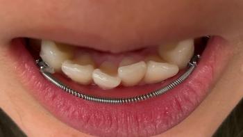
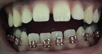
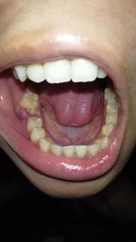
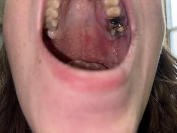
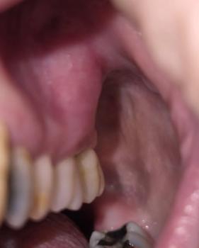
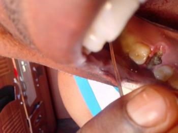
Patients with lower front gum inflammation may also experience tartar buildup, periodontal disease, gum abscesses, or tooth decay. Explore these related clinical case studies.
 |
 |
 |
 |
 |
 |