Smile Again with Comfort and Confidence.

Why Dentists Use Dental X-Rays: A Complete Guide to Diagnosis, Treatment Planning, and Patient Safety
image hero:
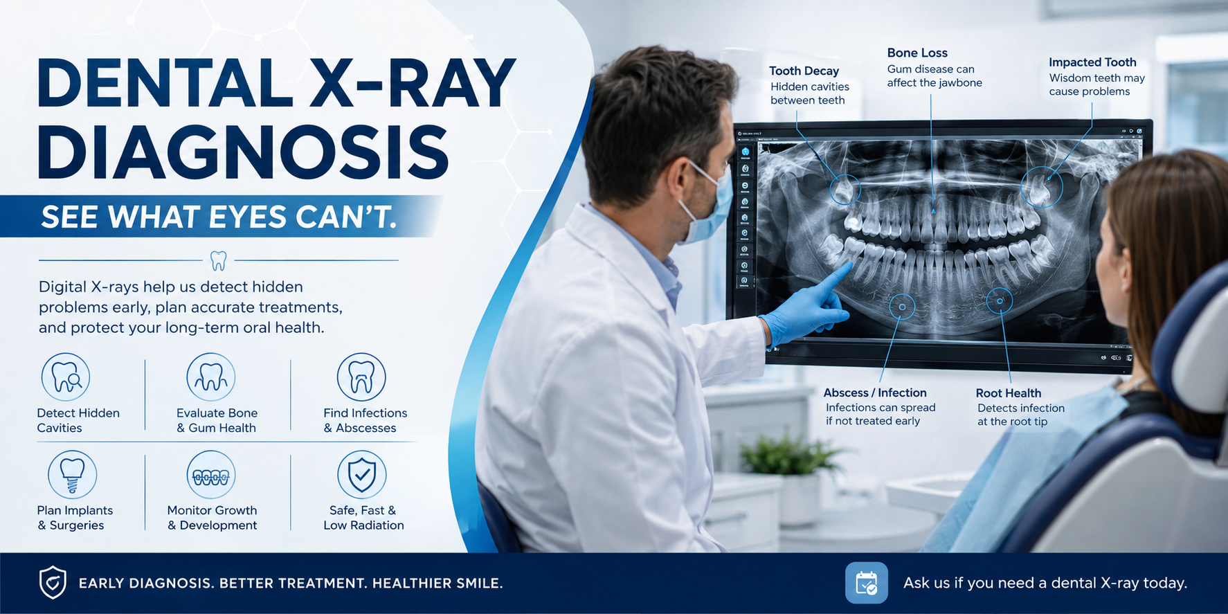
Dental X-rays (radiographs) are among the most valuable diagnostic tools in modern dentistry. While a clinical examination allows your dentist to inspect the visible surfaces of your teeth, gums, tongue, and oral tissues, many dental problems develop beneath the surface where they cannot be seen with the naked eye.
Digital dental X-rays provide detailed images of your teeth, roots, jawbone, and surrounding structures, allowing dentists to detect hidden conditions before they become painful, expensive, or difficult to treat. Early diagnosis often means simpler, more conservative treatment and a better long-term outcome.
Why a Visual Examination Alone Isn't Enough
Even the most experienced dentist cannot directly see:
- Tooth roots hidden beneath the gums
- Decay between tightly contacting teeth
- Bone supporting the teeth
- Developing infections inside the jawbone
- Impacted teeth
- Bone cysts or tumors
- Cracks extending below the gumline
- Internal tooth damage
- Changes around previous dental work
Dental X-rays allow clinicians to "see beyond the surface," making diagnosis significantly more accurate.
Key Reasons Dentists Use Dental X-Rays
1. Detecting Early Cavities Between Teeth
One of the most common reasons dentists recommend bitewing X-rays is to identify interproximal cavities—decay that forms between adjacent teeth.
These narrow contact areas cannot be fully examined during a visual inspection because:
- Toothbrush bristles cannot reach every surface
- The teeth block direct visualization
- Early cavities may not cause pain
- Enamel damage often begins beneath the surface
Without X-rays, these small cavities may continue growing unnoticed.
Early detection allows treatment with:
- Small tooth-colored fillings
- Minimal removal of healthy tooth structure
- Lower treatment costs
- Better long-term preservation of natural teeth
If left untreated, the decay may eventually reach the dental pulp, requiring:
- Root canal treatment
- Dental crown
- Tooth extraction
2. Identifying Hidden Decay Under Existing Fillings and Crowns
Dental restorations are durable but not permanent.
Over time, fillings, crowns, bridges, and veneers may:
- Wear down
- Crack
- Leak
- Separate from the tooth
Tiny openings may allow bacteria to enter beneath the restoration.
This recurrent decay is often invisible during a routine examination.
Dental X-rays help identify:
- Decay beneath fillings
- Marginal leakage
- Broken restorations
- Hidden fractures
- Secondary caries
Treating these problems early can prevent infection and preserve the tooth.
3. Evaluating Bone Loss from Gum Disease
Periodontal disease affects the bone supporting your teeth.
In its early stages, gum disease may produce only mild symptoms such as:
- Bleeding gums
- Swollen gums
- Bad breath
As the disease progresses, the jawbone gradually dissolves.
Dental X-rays allow dentists to evaluate:
- Bone height
- Bone density
- Vertical bone defects
- Horizontal bone loss
- Furcation involvement
- Periodontal pockets associated with bone destruction
This information is essential when planning:
- Scaling and root planing
- Periodontal maintenance
- Surgical periodontal treatment
- Long-term monitoring
4. Diagnosing Root Infections and Dental Abscesses
Many tooth infections develop silently.
The infection may begin after:
- Deep decay
- Dental trauma
- Cracked teeth
- Failed root canal treatment
An infected tooth may not produce immediate pain.
Dental X-rays can reveal:
- Periapical abscesses
- Chronic infections
- Granulomas
- Bone destruction
- Root resorption
- Hidden infection around tooth roots
Early diagnosis helps prevent infection from spreading into surrounding tissues.
5. Detecting Cysts, Tumors, and Other Jaw Abnormalities
Routine dental imaging occasionally identifies conditions unrelated to cavities.
Dentists may discover:
- Jaw cysts
- Benign tumors
- Impacted teeth
- Bone lesions
- Calcifications
- Developmental abnormalities
Many of these conditions produce no symptoms until they become large.
Finding them early greatly improves treatment outcomes.
6. Monitoring Children's Dental Development
Dental X-rays are especially valuable for growing children.
They help dentists monitor:
- Permanent tooth development
- Tooth eruption patterns
- Missing permanent teeth
- Extra teeth
- Impacted canines
- Wisdom tooth development
- Jaw growth
- Orthodontic spacing
Early detection allows orthodontic intervention before more complex problems develop.
7. Planning Dental Implant Treatment
Dental implants require careful planning.
Before implant placement, dentists must evaluate:
- Bone height
- Bone width
- Bone density
- Location of nerves
- Maxillary sinus position
- Existing pathology
- Adjacent tooth roots
Three-dimensional imaging helps determine the safest implant position while minimizing surgical risks.
8. Root Canal Treatment Planning
Root canal therapy depends on accurate imaging.
Dental X-rays help identify:
- Root length
- Number of canals
- Canal curvature
- Root fractures
- Periapical infection
- Previous treatment complications
Radiographs are also used during and after treatment to confirm successful cleaning and healing.
9. Planning Tooth Extractions
Not all teeth are straightforward to remove.
Before extraction, X-rays reveal:
- Root shape
- Root curvature
- Bone support
- Tooth position
- Relationship to nearby nerves
- Impacted wisdom teeth
This information reduces surgical complications and improves patient safety.
Common Types of Dental X-Rays
Bitewing X-Rays
Bitewing radiographs show the crowns of the upper and lower teeth together.
Best used for:
- Detecting cavities between teeth
- Monitoring existing fillings
- Evaluating early bone loss
- Routine preventive examinations
Periapical X-Rays (PA)
Periapical radiographs capture the entire tooth from crown to root tip.
Common uses include:
- Root infections
- Dental abscesses
- Root fractures
- Bone lesions
- Root canal treatment
Panoramic X-Rays
A panoramic X-ray captures the entire mouth in one image.
It includes:
- Upper jaw
- Lower jaw
- Wisdom teeth
- Jaw joints (TMJ)
- Maxillary sinuses
- Major nerve pathways
Common indications include:
- Wisdom tooth evaluation
- Orthodontic assessment
- General screening
- Impacted teeth
- Jaw abnormalities
Cone Beam CT (CBCT)
CBCT produces detailed three-dimensional images of hard tissues.
It is commonly recommended for:
- Dental implants
- Complex tooth extractions
- Root canal retreatment
- Impacted teeth
- Jaw pathology
- Bone grafting
- Oral surgery planning
Because CBCT provides much greater anatomical detail, it is generally prescribed only when clinically necessary.
Are Dental X-Rays Safe?
Yes. Modern digital dental X-rays are considered extremely safe when used appropriately.
Today's digital systems require significantly less radiation than traditional film-based radiography while producing clearer diagnostic images.
Dentists prescribe X-rays only when they provide important clinical information that cannot be obtained through a visual examination alone.
The ALARA Principle
Dentists in the United States and many other countries follow the ALARA Principle—As Low As Reasonably Achievable.
This means every X-ray examination is performed using the minimum amount of radiation necessary to obtain diagnostic-quality images.
Additional safety measures include:
- Digital sensors with lower radiation requirements
- Protective thyroid collars when appropriate
- Lead aprons when clinically indicated
- Limiting images to those that are medically necessary
- Individualized imaging schedules based on patient needs
American Dental Association (ADA) Recommendations
The American Dental Association recommends that dental X-rays be prescribed according to each patient's individual risk factors rather than on a fixed schedule.
Your dentist considers factors such as:
- Age
- History of cavities
- Existing restorations
- Gum disease
- Dry mouth
- Symptoms
- Previous dental treatment
- Overall oral health
Patients at higher risk for dental disease may benefit from more frequent imaging, while individuals with excellent oral health may require X-rays less often.
The Importance of Early Diagnosis
Many serious dental conditions begin without pain or visible symptoms.
By the time discomfort develops, treatment may become more complex and expensive.
Dental X-rays help detect problems while they are still small, allowing dentists to preserve more natural tooth structure and prevent avoidable complications.
When combined with regular dental examinations and good oral hygiene, dental radiographs play a vital role in maintaining healthy teeth, gums, and jawbone throughout life.
Related Clinical Cases
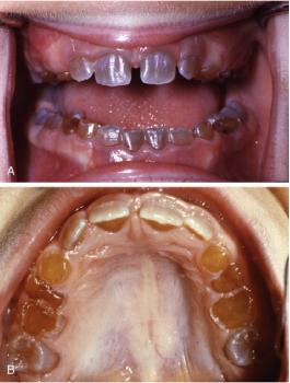
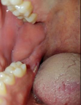
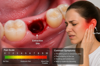
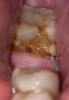
Patients with lower front gum inflammation may also experience tartar buildup, periodontal disease, gum abscesses, or tooth decay. Explore these related clinical case studies.



Other topics:
Other Dental Clinic