Smile Again with Comfort and Confidence.

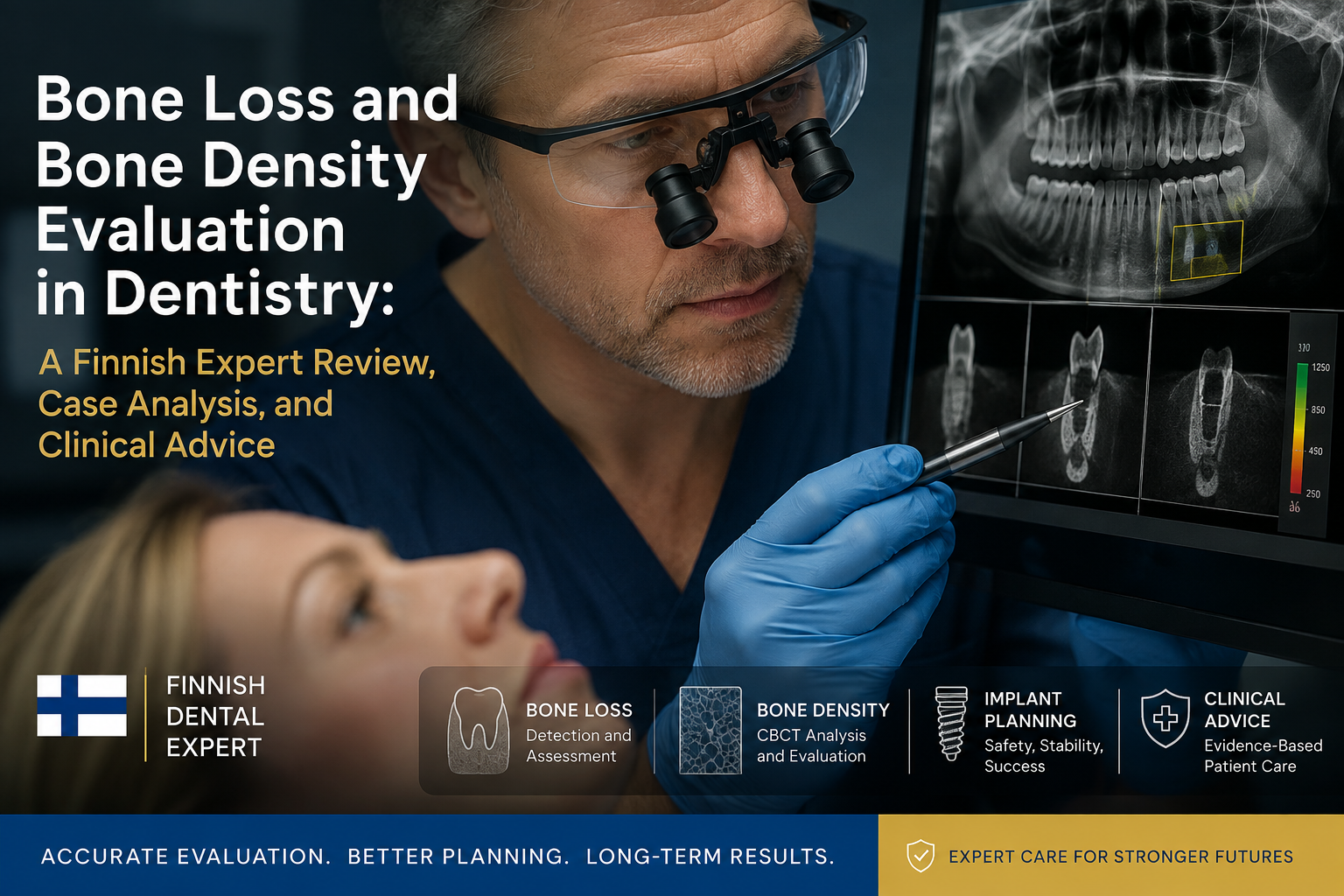
Bone Loss and Bone Density Evaluation in Dentistry: A Finnish Expert Review, Case Analysis, and Clinical Advice
image hero:
Clinical note: This article is an educational review written in the style of a Finnish dental expert. It is not a diagnosis of an individual patient. Actual treatment decisions require examination, appropriate imaging, and assessment by a qualified dentist or oral and maxillofacial specialist.
Introduction
Bone loss is one of the most important hidden problems in modern dentistry.
A patient may look healthy from the outside while significant loss of the supporting jawbone is already occurring beneath the gums. This becomes particularly important when a patient is considering dental implants, has advanced periodontal disease, has lost teeth, or has experienced long-term infection.
From a Finnish evidence-based dental perspective, bone should not be evaluated simply by asking whether there is "enough bone." The clinician should evaluate several characteristics:
-
Bone height
-
Bone width
-
Cortical bone thickness
-
Trabecular bone structure
-
Local bone density
-
Relationship to nerves and sinuses
-
Presence of infection or pathology
-
Periodontal condition
-
Previous tooth loss and extraction history
-
General skeletal health when clinically relevant
For implant treatment, three-dimensional imaging can provide valuable information about the anatomy of the proposed implant site. ITI consensus guidance identifies CBCT as the preferred cross-sectional imaging method when 3D assessment is indicated, while emphasizing that imaging should be justified according to the individual patient's clinical needs.
1. What Is Dental Bone Loss?
Dental bone loss refers primarily to the reduction of alveolar bone supporting the teeth.
The alveolar bone surrounds and supports the roots of the teeth. When the supporting tissues are damaged by periodontal disease, infection, trauma, or tooth loss, this bone can gradually disappear.
Bone loss can be:
Horizontal bone loss
The bone level decreases relatively evenly around a tooth or group of teeth.
Vertical bone loss
The bone disappears in a more localized, angular pattern, creating a deeper defect alongside the tooth.
Localized bone loss
Only one tooth or a small region is affected.
Generalized bone loss
Bone reduction occurs throughout much of the dentition.
Post-extraction bone loss
After a tooth is removed, the alveolar ridge commonly undergoes remodeling and reduction because the bone that supported the tooth is no longer receiving the same functional stimulation.
This is one reason why delaying tooth replacement for many years can complicate future implant treatment.
2. Bone Loss Is Not the Same as Low Bone Density
This distinction is extremely important.
A patient can have:
Adequate bone density but insufficient bone volume.
Or:
Adequate bone volume but relatively poor bone quality.
Or both problems simultaneously.
For example, a patient may have a wide ridge but low-density cancellous bone. Another patient may have dense cortical bone but a very narrow ridge.
Therefore, implant planning should not depend on a single number.
The clinician needs to evaluate the complete three-dimensional anatomy.
3. Why Bone Density Matters for Dental Implants
A dental implant must achieve sufficient primary stability at placement.
Primary stability is influenced by several factors, including:
-
Bone density
-
Bone volume
-
Implant geometry
-
Implant diameter
-
Implant length
-
Surgical technique
-
Preparation of the osteotomy
-
Cortical engagement
-
Implant position
Research has demonstrated a relationship between local bone density and implant stability. One study found a significant correlation between local bone density and implant stability, while patients with osteoporosis showed somewhat lower implant stability measurements than comparison groups.
Another clinical study found that alveolar bone density and ridge width were associated with primary implant stability.
However, bone density should not be interpreted as an isolated predictor of implant success.
4. How Is Bone Density Evaluated?
A dental clinician may use several complementary approaches.
Clinical examination
The dentist evaluates:
-
Gum health
-
Tooth mobility
-
Periodontal pockets
-
Recession
-
Previous tooth loss
-
Ridge contour
-
Signs of infection
-
Occlusion and bite forces
Clinical examination provides the foundation for deciding whether additional imaging is necessary.
Periapical radiographs
Small intraoral radiographs can provide valuable information about:
-
Crestal bone levels
-
Root anatomy
-
Periapical pathology
-
Periodontal bone changes
-
Existing restorations
They are particularly useful for localized assessment.
Panoramic radiography
A panoramic image provides a broad overview of:
-
Both jaws
-
Remaining teeth
-
Gross bone levels
-
Mandibular canal
-
Maxillary sinuses
-
Large pathological lesions
However, panoramic imaging does not provide the same three-dimensional information as CBCT.
5. CBCT Bone Evaluation
When three-dimensional assessment is clinically indicated, CBCT can provide cross-sectional information about the proposed implant region.
The clinician can assess:
Bone height
How much vertical bone is available?
Bone width
Is the ridge sufficiently wide?
Cortical boundaries
How thick and intact are the cortical plates?
Anatomical structures
Where are the mandibular canal, mental foramen, nasal cavity and maxillary sinus?
Pathology
Is there an unexpected lesion, infection, or other abnormality?
ITI consensus recommendations state that CBCT should be used when additional information beyond clinical examination and conventional radiography is needed. The field of view should be restricted to the relevant region whenever possible to reduce radiation exposure.
6. Can CBCT Measure Bone Density?
This requires an important clinical qualification.
CBCT images can produce voxel values that may be used to estimate bone density, and research has demonstrated associations between CBCT-derived density measurements and implant stability.
However, CBCT-derived Hounsfield-unit measurements should not automatically be treated as equivalent to standardized medical CT measurements.
Different CBCT machines, acquisition protocols, reconstruction methods and exposure parameters can influence voxel values.
A 2026 study specifically highlighted that conventional CBCT-derived HU measurements can be inaccurate and unreliable for quantitative bone-density assessment, with mixed evidence regarding their ability to predict implant primary stability.
Therefore, I would advise patients:
Do not make an implant decision based on a single CBCT density number.
The density measurement should be interpreted together with the patient's anatomy and clinical condition.
7. Case Analysis: The Patient With Long-Term Missing Tooth
Patient presentation
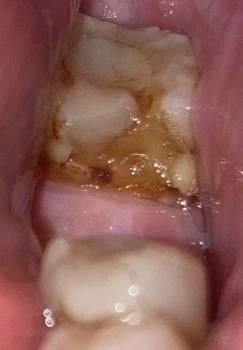
A hypothetical 58-year-old patient presents for replacement of a missing upper molar.
The tooth was extracted approximately eight years earlier.
The patient says:
"I was told I have enough bone because the area doesn't hurt."
This statement requires careful correction.
Clinical findings
The clinician observes:
-
Significant ridge narrowing
-
No acute pain
-
Healthy-looking mucosa
-
Reduced ridge width
-
Limited vertical bone beneath the maxillary sinus
A panoramic radiograph provides an initial overview.
CBCT is then considered because three-dimensional anatomy is necessary for implant planning.
CBCT assessment
The imaging demonstrates:
-
Reduced horizontal ridge width
-
Limited vertical implant space
-
Close proximity to the maxillary sinus
-
Relatively low-density posterior maxillary bone
The case illustrates an important principle:
Absence of pain does not mean absence of bone loss.
Bone deficiency can remain completely asymptomatic.
8. Clinical Interpretation of the Case
The implant clinician now has several possible strategies.
Depending on the patient's anatomy, periodontal status and treatment objectives, possibilities may include:
Option 1: Implant placement without augmentation
This may be appropriate when sufficient bone exists for predictable implant positioning.
Option 2: Guided bone regeneration
Bone augmentation may be considered when ridge width or contour is inadequate.
Option 3: Sinus augmentation
In selected posterior maxillary cases, additional vertical bone may be created through sinus augmentation.
Option 4: Alternative implant design or position
Sometimes the treatment plan can be modified to take advantage of the existing anatomy.
Option 5: Non-implant restoration
An implant is not automatically the correct treatment.
A bridge, removable prosthesis, or other restorative solution may sometimes be more appropriate.
9. What Does "Poor Bone Quality" Mean?
Dentists commonly describe bone as relatively dense or porous.
A simplified clinical concept is:
Dense cortical bone
Provides strong mechanical support and can contribute substantially to primary implant stability.
Trabecular or cancellous bone
Is more porous and has a different mechanical structure.
The posterior maxilla often presents a more challenging bone environment than the anterior mandible. Research has demonstrated substantial regional variation in jawbone density, with posterior maxillary sites commonly showing lower-density bone.
But again, these are general patterns—not a diagnosis for an individual patient.
10. Bone Density and Osteoporosis
This is another area where patients frequently become confused.
Osteoporosis affects the skeleton and can potentially influence the bone environment relevant to dental treatment.
Research has found associations between skeletal/local bone density and implant stability.
However:
Having osteoporosis does not automatically mean a patient cannot receive dental implants.
The dentist should instead consider:
-
Osteoporosis diagnosis
-
Fracture history
-
Medication history
-
Overall health
-
Local jawbone anatomy
-
Implant stability requirements
-
Healing considerations
-
Coordination with the patient's physician when necessary
A patient taking antiresorptive medication should specifically inform the dentist before invasive dental surgery.
11. Bone Loss From Periodontal Disease
Periodontitis is one of the major causes of tooth-supporting bone destruction.
The disease can progressively damage:
-
Gingival tissues
-
Periodontal ligament
-
Alveolar bone
As bone disappears around teeth, tooth mobility can develop.
Advanced periodontal disease may eventually result in tooth loss.
This creates a difficult cycle:
Periodontal disease → bone loss → tooth loss → ridge remodeling → more complicated tooth replacement
Therefore, periodontal disease must be controlled before implant placement.
An implant placed into an untreated inflammatory environment is not a substitute for periodontal treatment.
12. The Infection Question
Bone loss may also occur around infected teeth.
A chronic periapical infection can destroy surrounding bone.
Radiographically, this may appear as a radiolucent lesion around the root apex.
The treatment may involve:
-
Root canal treatment
-
Retreatment
-
Extraction
-
Removal of infected tissue
-
Bone healing
-
Later implant assessment
The important principle is:
Treat the disease before rebuilding the structure.
A dental implant should not simply be placed into an untreated infected site without appropriate clinical management.
13. Reviews: What I Would Look For Before Implant Treatment
From an evidence-based Finnish clinical perspective, I would review the case in five stages.
Review 1: Medical history
Check:
-
Systemic diseases
-
Osteoporosis
-
Medications
-
Smoking
-
Previous radiation therapy
-
Healing problems
-
Relevant surgical history
Review 2: Periodontal status
Assess:
-
Pocket depths
-
Bleeding
-
Tooth mobility
-
Plaque control
-
Existing periodontal destruction
Review 3: Radiographic anatomy
Evaluate:
-
Bone height
-
Bone width
-
Cortical plates
-
Anatomical boundaries
-
Pathology
Review 4: Implant biomechanics
Consider:
-
Implant diameter
-
Implant length
-
Implant position
-
Primary stability
-
Loading strategy
Review 5: Long-term maintenance
The implant is not the end of treatment.
The patient must maintain:
-
Excellent oral hygiene
-
Professional maintenance
-
Periodontal health
-
Regular implant monitoring
-
Control of excessive occlusal forces
14. Important Advice for Patients
If your dentist tells you that you have bone loss, do not immediately assume that dental implants are impossible.
Instead, ask:
1. How much bone have I lost?
Ask about both height and width.
2. Is the bone loss localized or generalized?
This can help identify whether the problem is associated with one tooth or broader periodontal disease.
3. Is the bone dense enough for the planned implant?
Ask how the clinician assessed bone quality.
4. Do I need CBCT?
CBCT should be clinically justified rather than performed automatically.
5. Is there active infection?
Any infection should be properly evaluated before implant surgery.
6. Do I need bone augmentation?
Ask why augmentation is recommended and what alternative treatments exist.
7. Do I have periodontal disease?
If periodontal disease is active, it needs appropriate treatment and control.
8. Are my medications important?
Tell the dentist about all prescription medications, particularly medications affecting bone metabolism.
15. My Finnish-Style Clinical Advice
A Scandinavian approach to dentistry generally emphasizes prevention, evidence-based decision-making, conservative treatment when appropriate, and long-term oral health rather than simply completing a procedure quickly.
For bone-loss cases, my clinical philosophy would be:
Do not rush.
Bone augmentation and implant surgery should be planned carefully.
Diagnose the cause.
Bone loss is a finding, not the final diagnosis.
Measure the anatomy.
Do not rely solely on visual inspection or a panoramic image when three-dimensional information is necessary.
Treat inflammation.
Periodontal disease and infection must be addressed.
Evaluate the whole patient.
Local bone anatomy is important, but systemic health and medication history also matter.
Plan for the next decade, not just the next appointment.
A successful implant should be designed for long-term maintenance.
16. What Patients Should Not Assume
Myth: "I have bone loss, so I cannot have implants."
Not necessarily.
Some patients can receive implants after appropriate periodontal treatment or bone augmentation.
Myth: "My CBCT says 700 HU, so my implant will definitely succeed."
No.
Bone-density measurements are only one part of treatment planning, and CBCT-derived density values have technical limitations.
Myth: "My jaw doesn't hurt, so my bone is healthy."
Not necessarily.
Periodontal bone loss and chronic dental disease can be clinically silent.
Myth: "Bone grafting guarantees implant success."
No procedure provides a guarantee.
Outcome depends on patient factors, anatomy, surgical technique, healing, restoration and long-term maintenance.
17. Dental Hub Clinical Summary
Bone loss should be evaluated as a three-dimensional biological and mechanical problem.
For implant dentistry, the clinician should consider:
Bone volume + bone quality + anatomy + infection status + periodontal health + systemic factors + implant design + surgical strategy.
CBCT is an important tool when 3D assessment is justified, particularly for evaluating implant-site anatomy. ITI consensus guidance supports CBCT for situations where conventional examination and imaging do not provide sufficient information.
At the same time, modern evidence cautions against treating CBCT-derived density numbers as perfectly standardized measurements of bone mineral density.
The best implant plan is therefore not:
"Find a high-density number and place an implant."
It is:
Diagnose → Measure → Control disease → Plan anatomy → Select treatment → Confirm stability → Maintain long term.
Final Dental Expert Advice
If you have been told that you have bone loss or low bone density, ask your dentist for a complete explanation of:
-
Where the bone has been lost
-
Why the bone has been lost
-
Whether infection or periodontal disease is present
-
How much bone remains
-
Whether CBCT is clinically indicated
-
Whether bone augmentation is necessary
-
Whether your systemic health or medications affect treatment
-
What implant and non-implant alternatives are available
-
How the proposed treatment will be maintained long term
A sophisticated dental treatment plan does not begin with the implant.
It begins with understanding the bone.
Evidence Review
The clinical principles in this review are consistent with published research and international implantology consensus guidance concerning CBCT, implant-site anatomy, bone density and primary implant stability.
Dental Hub Editorial Position: Bone density should be interpreted as one component of comprehensive implant assessment rather than as a standalone predictor of treatment success.
Related Clinical Cases
Patients with lower front gum inflammation may also experience tartar buildup, periodontal disease, gum abscesses, or tooth decay. Explore these related clinical case studies.



Other topics:
Other Dental Clinic