Modern Dentistry, Timeless Smiles.

Severe Lower Molar Decay: Symptoms, Root Canal, Treatment Options, Recovery & When It's a Dental Emergency
Image:
Severity:
Teeth Problems:
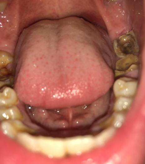
Severe Lower Molar Decay: Urgent Restoration and Gum Care Needed
Case Type: Advanced Dental Caries and Calculus Accumulation
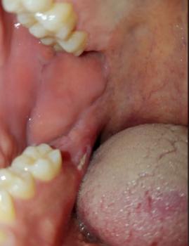
A 42-year-old patient presented with severe pain while chewing on the lower right molar. Clinical examination revealed extensive occlusal and proximal decay with bleeding gums and deep periodontal pocketing. Bitewing and periapical radiographs showed decay extending close to the pulp while moderate bone loss surrounded the tooth. After periodontal therapy and root canal treatment, the tooth was restored with a zirconia crown, allowing the patient to regain normal chewing function.
Full Analysis and Diagnosis
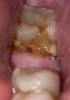
Upon examination, the posterior molars exhibit:
-
Extensive caries (tooth decay) on both left and right sides.
-
Possible pulp involvement — dark discoloration and cavity penetration indicate the decay may have reached the pulp chamber.
-
Heavy calculus deposits and plaque buildup along the gumline, likely contributing to inflammation or gingivitis.
-
Gingival swelling may be present, suggesting bacterial infection and poor oral hygiene.
Recommended Dental Procedures
-
Initial Phase (Day 1–3):
-
Full oral cleaning and scaling to remove calculus and bacterial plaque.
-
Caries excavation and temporary filling to prevent further infection.
-
-
Intermediate Phase (Day 4–10):
-
Endodontic evaluation (root canal treatment) if pulpal infection or pain is present.
-
Restoration or extraction depending on tooth structure integrity.
-
-
Final Phase (Day 11–14):
-
Permanent fillings or crowns for restored teeth.
-
Soft tissue therapy (antiseptic rinses, possible antibiotics if infection persists).
-
Why Lower Molars Fail More Often
- Deep pits and fissures
- High chewing forces
- Difficult access for brushing
- Food impaction
- Multi-root anatomy
- Plaque retention
- Gum inflammation
Severe Lower Molar Decay: Why Immediate Treatment Matters
Lower molars are among the hardest-working teeth in your mouth. Every meal places tremendous chewing pressure on these teeth. Unfortunately, their deep grooves and location near the back of the mouth also make them one of the most common places for severe tooth decay to develop.
When decay is ignored, bacteria can destroy enamel, penetrate dentin, infect the dental pulp, inflame surrounding gums, and eventually spread into the jawbone. At this stage, treatment becomes significantly more complex—and delaying care can increase the risk of losing the tooth or developing a serious infection.
Clinical Case Study
Patient Overview
Age: 42 years old
Chief Complaint
The patient reported:
- Severe pain while chewing
- Lingering sensitivity to hot and cold beverages
- Bleeding gums around the lower molar
- Bad taste in the mouth
- Difficulty chewing on one side
The discomfort had gradually worsened over several months before becoming severe enough to seek dental treatment.
Clinical Examination
The dental examination revealed:
- Extensive decay involving the chewing surface and side of a lower mandibular molar
- Significant loss of enamel
- Deep dentin destruction
- Gingival inflammation surrounding the tooth
- Bleeding during periodontal probing
- Deep periodontal pocket adjacent to the affected molar
- Heavy plaque accumulation near the gumline
The remaining tooth structure appeared weakened and at risk for fracture.
Radiographic Findings
Periapical and bitewing radiographs demonstrated:
- Deep decay approaching the pulp chamber
- Extensive dentin destruction
- Bone loss adjacent to the infected tooth
- Localized periodontal involvement
- Multi-rooted anatomy requiring careful treatment planning
Fortunately, the tooth still possessed enough remaining structure to be considered potentially restorable.
Clinical Diagnosis
The patient was diagnosed with:
- Severe dental caries involving the lower mandibular molar
- Advanced dentin destruction
- Symptomatic irreversible pulpitis (or pulpal involvement, depending on vitality testing)
- Localized chronic periodontitis surrounding the tooth
- Gingival inflammation with bleeding on probing
Without prompt treatment, the tooth was at significant risk for abscess formation, root fracture, and eventual extraction.
Why Lower Molars Are Especially Vulnerable
Lower molars are naturally more susceptible to advanced decay because they contain:
- Deep pits and fissures
- Narrow grooves that trap bacteria
- Broad chewing surfaces
- Tight contact points between teeth
- Multiple roots that complicate cleaning
Since these teeth perform the majority of chewing, they experience constant mechanical stress. Food particles and bacterial plaque often remain trapped in difficult-to-clean areas, especially when flossing is inconsistent.
Over time, bacteria convert sugars into acids that dissolve enamel. As decay progresses into dentin, bacteria multiply rapidly and move toward the nerve.
At the same time, plaque accumulating beneath the gumline triggers chronic inflammation, causing swollen gums, bleeding, and periodontal pocket formation.
How Tooth Decay Progresses
Stage 1: Enamel Demineralization
Acids produced by oral bacteria begin dissolving minerals from the enamel.
Symptoms are usually absent.
Stage 2: Dentin Decay
Once bacteria penetrate dentin:
- Cavities enlarge rapidly
- Food becomes trapped
- Cold sensitivity develops
- Chewing discomfort begins
Stage 3: Pulp Inflammation
When bacteria reach the pulp:
- Severe spontaneous toothache develops
- Hot and cold sensitivity lingers
- Nighttime pain becomes common
- Inflammation affects blood vessels and nerves
Stage 4: Infection Beyond the Tooth
If untreated:
- The pulp dies
- Bacteria spread through root canals
- Infection reaches surrounding bone
- Dental abscess forms
- Facial swelling may develop
Periodontal Disease Makes the Situation Worse
When gum disease occurs alongside severe decay, treatment becomes more challenging.
Periodontal infection causes:
- Gum recession
- Bone loss
- Deeper periodontal pockets
- Tooth mobility
- Reduced long-term stability
Saving the tooth requires addressing both the cavity and the surrounding periodontal infection.
Comprehensive Treatment Plan
A multidisciplinary approach was recommended.
Step 1: Infection Control
Professional scaling and root planing removed plaque and hardened calculus beneath the gums.
This reduced bacterial load and allowed inflamed tissues to begin healing.
Step 2: Root Canal Therapy
Because the decay had reached the pulp:
- Infected nerve tissue was removed.
- Root canals were disinfected.
- The canals were sealed to prevent reinfection.
This eliminated pain while preserving the natural roots.
Step 3: Crown Lengthening (When Necessary)
If decay extends below the gumline, additional healthy tooth structure may need to be exposed surgically.
Crown lengthening creates a stable foundation for a long-lasting restoration.
Step 4: Full-Coverage Crown
After healing, a custom porcelain or zirconia crown was placed.
The crown restores:
- Strength
- Function
- Appearance
- Protection against fracture
Lower molars experience tremendous chewing forces, making full coverage essential after extensive tooth destruction.
Recovery Expectations
Following treatment, most patients experience gradual improvement over several days.
Temporary tenderness during chewing may occur but generally resolves as tissues heal.
Long-term success depends on maintaining excellent oral hygiene and attending regular dental examinations.
Prevention Tips
You can greatly reduce the risk of severe lower molar decay by:
- Brushing twice daily with fluoride toothpaste
- Cleaning between teeth every day with floss or interdental brushes
- Limiting sugary snacks and beverages
- Drinking plenty of water
- Receiving professional dental cleanings every six months
- Treating small cavities before they become large ones
- Considering dental sealants for deep grooves when appropriate
Frequently Asked Questions (FAQ)
Why are lower molars particularly vulnerable to severe decay and gum inflammation?
Lower mandibular molars contain deep pits, grooves, and tight contact areas that trap plaque and food particles. Because these teeth are located at the back of the mouth, they are often more difficult to clean thoroughly. Plaque left near the gumline produces acids that dissolve enamel while simultaneously triggering inflammation in the surrounding gum tissue, leading to cavities and periodontal disease occurring together.
When does severe lower molar decay become an urgent dental condition?
Immediate evaluation is recommended when:
- Pain becomes spontaneous or throbbing
- Sensitivity to hot or cold lingers
- Decay extends beneath the gumline
- Swelling develops
- Pus drains from the gums
- A bad taste persists
- Chewing becomes extremely painful
- An abscess forms
- The tooth begins breaking apart
These signs may indicate pulp infection or spread into surrounding tissues.
What diagnostic tests determine whether the tooth can be saved?
Your dentist may perform:
- Comprehensive clinical examination
- Periapical radiographs
- Bitewing radiographs
- Pulp vitality testing
- Percussion testing
- Bite testing
- Periodontal probing
- Mobility assessment
- Evaluation of furcation involvement
- Occlusal analysis
These findings help determine whether restoration, root canal therapy, periodontal treatment, or extraction offers the best long-term outcome.
What treatments are usually needed?
Depending on the severity, treatment may include:
- Scaling and root planing
- Antibiotics (when clinically indicated—not routinely for every cavity)
- Root canal therapy
- Crown lengthening surgery
- Core build-up
- Porcelain or zirconia crown
- Periodontal maintenance therapy
If the tooth cannot be predictably restored because of extensive structural destruction or severe bone loss, extraction followed by tooth replacement options such as a dental implant, bridge, or removable prosthesis may be recommended.
How should I care for the tooth during healing?
Follow your dentist's instructions carefully.
General recommendations include:
- Brush gently using a soft-bristled toothbrush.
- Clean between teeth daily.
- Use any prescribed mouth rinse exactly as directed.
- Eat softer foods until chewing is comfortable.
- Avoid chewing hard foods on the treated tooth.
- Attend all follow-up appointments.
Cold Compress Guidance
For mild swelling or discomfort:
- Apply a cold compress to the outside of the cheek for 10–20 minutes at a time during the first day if recommended by your clinician.
Avoid Heat
Do not apply warm compresses to a swollen face or jaw when an active dental infection is suspected. Heat may increase blood flow and can worsen swelling in some infections. Prompt dental evaluation is far more important than relying on home remedies.
Never Attempt Home Drainage
Do not squeeze, puncture, or attempt to drain swollen gums yourself. This can spread bacteria deeper into surrounding tissues and increase the risk of serious infection.
Emergency Warning: When a Dental Infection Becomes Life-Threatening
A localized tooth infection can occasionally spread beyond the tooth into the soft tissues of the face and neck. This may progress to facial cellulitis or a deep neck infection, both of which require urgent medical attention.
Go to the nearest emergency department or call emergency services immediately if you develop:
- Difficulty breathing
- Difficulty swallowing
- Excessive drooling or inability to swallow saliva
- Rapidly spreading swelling of the face, jaw, or neck
- Swelling extending toward the eye or chest
- High fever
- Chills
- Severe weakness or extreme fatigue
- Confusion or altered mental status
- A stiff neck accompanied by signs of infection
These symptoms may indicate that the infection is spreading beyond the tooth and requires emergency medical care.
Key Takeaway
Severe lower molar decay is more than just a cavity—it can involve the tooth, gums, supporting bone, and, in advanced cases, the surrounding facial tissues. Early diagnosis, appropriate restorative and periodontal treatment, and consistent oral hygiene offer the best chance of saving the tooth and preventing serious complications. If you experience worsening pain, swelling, or any emergency warning signs, seek prompt evaluation by a dentist or emergency medical services without delay.
Possible Complications if Untreated
-
Spread of infection to adjacent teeth and gums.
-
Tooth loss due to root destruction.
-
Jawbone infection (osteomyelitis) in severe untreated cases.
-
Chronic bad breath and systemic bacterial spread.
Expert Comment
“Early treatment is key. Even advanced decay can be saved with root canal therapy and restoration if handled promptly.”
Advice to the Patient
Visit your nearest dental clinic or implant specialist for immediate evaluation and treatment. Delaying care will only increase discomfort and treatment costs.
Search nearby dental experts here:
https://cebudentalimplants.com/map-dental-clinic
Related Clinical Cases
Patients with lower front gum inflammation may also experience tartar buildup, periodontal disease, gum abscesses, or tooth decay. Explore these related clinical case studies.

Other topics:
Other Dental Clinic