Where Bright Smiles Begin.

Heavy Dental Calculus and Gingival Inflammation: Comprehensive Clinical Assessment, Treatment Timeline, and 14-Day Healing Guide
Image:
Severity:
Teeth Problems:
A dentist would need periodontal probing, bite/occlusal examination, and usually appropriate radiographs to determine bone loss and whether there is an underlying dental infection.
What I can see at 100% view
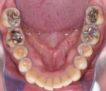
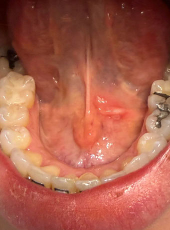
The most obvious finding is substantial hardened calculus/tartar on the tongue-side (lingual) surfaces of the lower front teeth, particularly around the lower incisors. There also appears to be localized redness/inflammation of the soft tissue under the tongue, near the floor-of-mouth/frenulum region.
The image does not by itself establish the severity of periodontal bone loss.
1. Heavy calculus — HIGH confidence
The yellow/brown deposits around the lower anterior teeth look consistent with mature dental calculus rather than simply soft plaque.
This is important because calculus provides a rough surface where plaque can accumulate and can contribute to gingival inflammation.
2. Gingival inflammation — likely
The surrounding gingival tissues appear somewhat erythematous. This could be compatible with gingivitis, but if periodontal pockets and bone loss are present, it could represent periodontitis.
A photograph cannot distinguish those reliably.
3. Floor-of-mouth lesion — needs attention
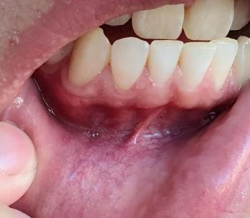
There is a localized reddish/elevated area beneath the tongue, close to the lingual frenulum.
This could have relatively benign explanations, including irritation or inflammation around normal salivary-duct structures, but the photograph alone cannot determine what it is.
This is the part I would specifically have a dentist/oral-medicine clinician examine.
What I would do clinically
Step 1 — Comprehensive examination
A dentist should document:
- Full periodontal charting
- Pocket depths around every tooth
- Bleeding on probing
- Tooth mobility
- Recession
- Furcation involvement where applicable
- Plaque/calculus levels
- Condition of existing restorations
- Examination of the entire floor of mouth and tongue
- Palpation of the floor of mouth and neck lymph nodes
The ADA recommends a conventional visual and tactile examination for oral mucosal abnormalities rather than relying on photographs alone.
Step 2 — Radiographs if clinically indicated
Depending on the periodontal findings, the dentist may obtain appropriate intraoral radiographs to determine whether there is:
- Horizontal bone loss
- Vertical bone defects
- Calculus below the gumline
- Root abnormalities
- Periapical pathology
- Other hidden dental disease
Radiographs can provide information that simply cannot be obtained from this photograph.
Step 3 — Professional scaling
For the visible calculus, professional removal is appropriate.
If there is only supragingival calculus, routine scaling may be sufficient.
If there is substantial subgingival calculus and periodontal pocketing, the treatment may instead require scaling and root planing/deep periodontal instrumentation, often divided into sections depending on the patient's findings.
If you want everything scaled
I would not recommend trying to remove this amount of calculus yourself with a metal scaler.
A reasonable professional sequence is:
Visit 1
→ Examination + periodontal charting
→ Determine whether radiographs are needed
→ Remove obvious supragingival calculus where appropriate
→ Establish periodontal diagnosis
Visit 2 or subsequent visits
→ Complete periodontal instrumentation/scaling
→ Root-surface debridement where indicated
→ Oral-hygiene instruction
Re-evaluation
→ Check bleeding, inflammation, pocket measurements and tissue response
→ Determine whether additional periodontal treatment is necessary.
What could improve within 14 days?
If the main problem is plaque/calculus-associated gingival inflammation and the irritant is professionally removed, the soft-tissue inflammation can begin improving within days.
However, 14 days does not guarantee complete healing.
A useful way to think about it:
| Finding | Possible response |
|---|---|
| Soft plaque | Improves quickly with effective cleaning |
| Gingival inflammation | Often noticeably improves over 1–2 weeks |
| Heavy calculus | Requires professional removal |
| Deep periodontal pockets | May require additional periodontal treatment |
| Bone loss | Does not simply regenerate in 14 days |
| Local traumatic irritation | May improve within days after the irritant is removed |
| Persistent unexplained mucosal lesion | Requires professional reassessment |
The important 14-day issue
The floor-of-mouth/red area is the finding I would not simply ignore for 14 days.
If that area is new and clearly attributable to irritation, the dentist may choose to remove the suspected irritant and observe the healing response.
But if a red/white lesion, ulcer, lump, thickening, or other unexplained abnormality persists for approximately two weeks, it should be reevaluated. The ADA states that persistent oral abnormalities warrant further investigation and, when appropriate, biopsy/referral.
That does NOT mean this photograph shows cancer. There are many benign causes of oral lesions. It means persistence beyond the expected healing period changes the clinical approach.
What could "scale up" if treatment is delayed?
If the calculus and inflammation are left untreated, the concern is progression from superficial inflammation to periodontal disease in susceptible patients:
Plaque → calculus → gingival inflammation → periodontal pocket formation → attachment loss → bone loss → tooth mobility/tooth loss
But that progression is not inevitable and cannot be predicted from this photograph alone.
Also, antibiotics are not a substitute for removing the dental cause when the problem is localized periodontal/dental disease. ADA guidance emphasizes definitive dental treatment rather than routine antibiotics for most localized dental conditions.
Seek prompt examination sooner than 14 days if
The floor-of-mouth area develops:
- Rapidly increasing swelling
- Significant pain
- Pus
- Fever
- Difficulty swallowing
- Difficulty breathing
- Difficulty moving the tongue
- Neck swelling
- Rapidly worsening symptoms
Those symptoms require urgent clinical assessment.
My overall impression from this image: the heavy lower-anterior calculus is the clearest dental problem, with probable localized gingival inflammation. The under-tongue red/elevated area deserves a separate examination, because its nature cannot be reliably determined from this photograph.
If this is being prepared as a CebuDentalImplants.com dental case, I would label it “Preliminary photographic assessment — not a definitive diagnosis” and recommend periodontal charting + appropriate radiographs + examination of the floor of mouth before assigning the final diagnosis.
Related Clinical Cases
Patients with lower front gum inflammation may also experience tartar buildup, periodontal disease, gum abscesses, or tooth decay. Explore these related clinical case studies.