Missing Teeth? Find Your Smile Again.

Interproximal Tooth Decay: How Dental X-Rays Help Detect Hidden Cavities Between Teeth
Image:
Severity:
Teeth Problems:
Why Cavities Between Teeth Need Special Attention
Interproximal tooth decay develops on the surfaces where two neighboring teeth touch. These areas are difficult to inspect directly, especially between the back molars and premolars. Food debris and dental plaque can accumulate in these spaces, increasing the risk of tooth decay when oral hygiene and fluoride protection are inadequate.
As a dental health educator, one important point to understand is that a tooth can have decay even when it looks normal from the outside and does not hurt. Early cavities may have few or no symptoms, making professional examination particularly important.
Bitewing dental X-rays can help dentists detect decay between posterior teeth, estimate how deeply a lesion extends, and determine whether further evaluation or treatment is needed. However, an X-ray finding must be interpreted alongside the patient's symptoms, clinical examination, and individual risk factors.
How Dentists Interpret an Interproximal Tooth Decay X-Ray
When examining a dental radiograph, a dentist looks for changes in the density of the tooth structure, including darker areas that may indicate mineral loss.
Potential findings include:
-
Early enamel demineralization: A small radiolucent area may suggest mineral loss within the enamel.
-
Decay extending into dentin: A lesion extending beyond the enamel may require closer assessment and possibly restorative treatment.
-
Possible cavitation: The dentist evaluates whether the tooth surface has developed an actual cavity. An X-ray alone cannot always establish whether the surface has broken down.
-
Decay near an existing filling: Recurrent decay may develop next to an older restoration, although a dark line near a filling does not automatically prove that the restoration has failed.
-
Changes in supporting bone: Bone-level changes may suggest periodontal disease or another condition, but they require separate clinical assessment and should not automatically be attributed to tooth decay.
The depth and activity of a lesion matter more than its appearance alone. A dentist may compare current and previous radiographs, inspect the tooth, evaluate sensitivity, and assess the patient's cavity risk before recommending treatment.
Does Dental Scaling Remove Cavities Between Teeth?
No. Professional scaling removes plaque and hardened tartar, but it does not repair a cavity or restore lost tooth structure.
Scaling may be recommended when plaque and calculus contribute to gum inflammation, bleeding, or periodontal disease. It helps improve the health of the surrounding gum tissues, but the tooth decay must be assessed and managed separately.
Depending on the clinical findings, treatment may include:
-
Fluoride treatment and monitoring: Appropriate for selected early, noncavitated lesions that may be managed without drilling.
-
A dental filling: Often used when a cavity has formed and requires restoration.
-
Replacement of a defective restoration: Considered when an existing filling has failed or recurrent decay is confirmed.
-
Root canal treatment: May be necessary when the pulp inside the tooth is irreversibly inflamed or infected and the tooth can be preserved.
-
A crown or other protective restoration: May be appropriate when substantial tooth structure has been lost.
-
Tooth extraction: Considered when the tooth cannot be predictably restored or saved.
The correct treatment cannot be selected from the darkness of an X-ray alone. A dentist must evaluate the tooth's structure, pulp condition, restorability, and surrounding tissues.
What Happens if Interproximal Tooth Decay Is Ignored?
Untreated active decay may gradually progress deeper into the tooth. Depending on its location and severity, possible complications include:
-
Breakdown of enamel and dentin.
-
Food trapping and increasing sensitivity.
-
Inflammation of the dental pulp.
-
Pulp infection or death of the pulp.
-
Infection around the root, including a possible dental abscess.
-
Extensive tooth damage that makes treatment more complex.
Progression is not identical in every patient. Some early lesions remain stable or can be managed with preventive care, while others progress. Regular examinations and individualized monitoring help dentists decide when treatment is necessary.
A dental abscess may cause severe pain, swelling, pus, or fever. Facial or neck swelling, difficulty swallowing, or difficulty breathing requires urgent medical or dental assessment; breathing or swallowing difficulty is an emergency.
Daily Prevention: Protecting the Spaces Between Teeth
A practical prevention routine includes the following:
-
Brush twice daily with fluoride toothpaste.
-
Clean between the teeth every day using dental floss or a suitable interdental brush.
-
Reduce frequent consumption of sugary snacks and sweetened drinks.
-
Drink water regularly, particularly after meals.
-
Attend dental examinations according to your dentist's recommended schedule.
-
Have dental X-rays when clinically indicated, rather than following an arbitrary schedule.
Flossing helps remove plaque from contact areas that a toothbrush may not reach effectively. However, flossing cannot repair an established cavity. If food repeatedly becomes trapped between the same teeth, ask your dentist to assess the contact area, restorations, and gum health.
Short Patient Story: A Teacher Discovers a Hidden Cavity
The following is a fictional educational case, not a real patient testimonial.
Maria was a 39-year-old public school teacher who spent most of her day preparing lessons, checking assignments, and helping students. Because she had no significant toothache, she postponed her routine dental appointment several times.
One afternoon, she noticed that food frequently became trapped between two lower molars. Occasionally, the gum in that area bled when she cleaned her teeth. She assumed that professional cleaning alone would solve the problem.
During her dental visit, the dentist examined her gums and teeth and recommended a bitewing X-ray because the contact area could not be adequately assessed visually. The image showed a suspicious area between the molars that required further clinical evaluation.
The dentist explained that scaling could remove tartar and help manage plaque-related gum inflammation, but it would not repair a confirmed cavity. After assessing the tooth and determining the extent of the lesion, the dentist recommended the appropriate treatment.
Maria also learned how to clean between her molars and reduce frequent sugary snacks during long teaching days. She arranged follow-up care around her school schedule instead of waiting for pain to develop.
The lesson: A lack of toothache does not guarantee that teeth are healthy. Early assessment can help patients understand their options before a small problem becomes more complicated.
When Should You See a Dentist?
Arrange a dental examination if you notice persistent food trapping, new sensitivity, pain when chewing, visible tooth damage, or gums that repeatedly bleed.
Seek prompt dental care for worsening tooth pain, swelling, or suspected infection. Fever accompanied by dental swelling requires urgent assessment, and difficulty breathing or swallowing warrants emergency care.
An X-ray is a diagnostic aid, not a complete diagnosis by itself. The dentist will determine which tests and treatments are appropriate for your individual situation.
Frequently Asked Questions About Interproximal Tooth Decay
1. Can a dental X-ray detect cavities between teeth?
Yes. Bitewing radiographs are commonly used to detect decay between posterior teeth when the contacting surfaces cannot be adequately inspected visually. Their ability to detect a lesion depends on its size, depth, location, and the quality of the image. A clinical examination is still necessary.
2. Can an interproximal cavity exist without tooth pain?
Yes. Early and even some more advanced cavities may not cause noticeable pain. Symptoms can develop when decay approaches or affects the pulp. Do not wait for a toothache before arranging an examination if your dentist has identified a suspicious lesion.
3. Can scaling cure interproximal tooth decay?
No. Scaling removes plaque and tartar, which can improve gum health, but it cannot replace missing enamel or dentin. A confirmed cavity requires a separate treatment decision based on its activity, depth, and whether the tooth surface has broken down.
4. Can early decay between teeth heal naturally?
An early, noncavitated lesion may be arrested or partially remineralized with appropriate fluoride exposure, plaque control, and dietary changes. Once a substantial cavity has formed, lost tooth structure generally cannot grow back, and a restoration may be necessary.
5. Does every dark spot on a dental X-ray mean a cavity?
No. Radiolucent areas may be associated with decay, but interpretation depends on the location, shape, image quality, and surrounding anatomy. Overlapping structures, restorative materials, and other factors can complicate interpretation. A dentist must correlate the image with clinical findings.
6. Will I need a root canal for a cavity between my molars?
Not necessarily. Many interproximal cavities can be treated with a filling. Root canal treatment may be required if the pulp is irreversibly inflamed or infected and the tooth is suitable for preservation. The depth of the lesion, symptoms, clinical tests, and radiographic findings guide this decision.
7. How can I prevent cavities between my teeth?
Brush twice daily with fluoride toothpaste, clean between teeth daily, limit frequent sugar exposure, and attend dental checkups at an interval appropriate for your risk level. Your dentist may recommend additional fluoride or other preventive measures if you are at increased risk of decay.
8. Are dental X-rays safe?
Dental X-rays use ionizing radiation, so they should be prescribed when the expected diagnostic benefit justifies the exposure. Modern dental imaging uses dose-reduction measures, and the appropriate imaging method depends on the clinical question, patient history, and individual needs. Tell your dentist if you are pregnant or may be pregnant so imaging decisions can be made appropriately.
9. What should I do if food keeps getting stuck between two teeth?
Clean the area gently with dental floss or an appropriately sized interdental brush. Avoid forcing objects into the gum. Persistent food trapping may be associated with a cavity, an open contact, gum recession, or a defective filling, so arrange an examination to identify the cause.
10. Can untreated tooth decay cause a dental abscess?
Yes. If decay progresses into the pulp and the infection spreads beyond the root, a dental abscess may develop. Symptoms can include severe pain, swelling, pus, and fever. A suspected abscess needs prompt professional treatment; antibiotics alone do not eliminate the underlying dental source.
Related Dental Reading
Final Clinical Advice
Interproximal tooth decay can be difficult to identify without a professional examination, and some lesions progress without obvious symptoms. Dental X-rays help dentists evaluate hidden areas, while scaling supports gum health when plaque and tartar are present. Neither a radiographic finding nor a cleaning appointment should replace an individualized treatment plan.
Medical disclaimer: This article provides general dental education and does not establish a diagnosis or replace an examination by a licensed dentist. Treatment recommendations depend on the patient's clinical findings, symptoms, medical history, and appropriate diagnostic imaging.
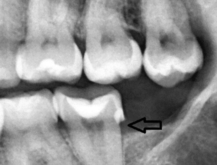
Teeth Case – Zoom 100% (Dental X-ray)
Focused Area: Posterior teeth (molar region) with interproximal defect indicated by arrow
Full Analysis and Deep Radiographic Examination
This assessment is based on the provided dental X-ray image only and does not replace a complete clinical examination, periodontal probing, or additional radiographs if required.
Radiographic Findings
-
Radiolucent area between posterior teeth, consistent with interproximal dental caries
-
Possible loss of tooth structure at the contact point
-
Early signs of localized bone level reduction adjacent to the affected tooth
-
Existing restoration visible on the molar, suggesting previous decay history
-
No obvious advanced periapical abscess visible at this image depth, but early pathology cannot be ruled out
Probable Diagnosis
-
Interproximal dental caries (moderate depth)
-
Early periodontal bone involvement localized to the molar region
-
Increased risk of pulp irritation depending on cavity depth
Recommended Treatment Process
Step 1: Full Mouth Scaling and Polishing (Day 1)
-
Ultrasonic scaling to remove plaque and calculus
-
Manual scaling in posterior and interproximal areas
-
Polishing to reduce bacterial reattachment
Estimated duration: 45–60 minutes
Local anesthesia: May be required depending on sensitivity
Step 2: Definitive Interproximal Treatment (Day 1–7)
Based on X-ray depth and clinical findings:
-
Composite restoration for moderate interproximal decay
-
Replacement of failing restoration if margins are compromised
-
Root canal treatment if pulp involvement is confirmed
-
Crown placement if structural integrity is reduced
Healing and Recovery Timeline
| Time Frame | Expected Outcome |
|---|---|
| Days 1–3 | Gum irritation decreases after scaling |
| Days 4–7 | Stabilization of gingival tissues |
| Days 8–14 | Healthy gum response if decay is treated |
A 14-day healing period is realistic for soft tissue once infection sources are removed.
What Will Scale Up If Left Untreated
-
Expansion of interproximal decay into the pulp
-
Development of dental abscess or chronic infection
-
Progressive bone loss around the affected molar
-
Increased tooth mobility and eventual tooth loss
-
Higher cost and complexity of future dental treatment
Post-Treatment Care Instructions
-
Brush twice daily using fluoride toothpaste
-
Floss daily, especially between molars
-
Use interdental brushes if contacts are open
-
Maintain regular dental checkups and X-rays as advised
Professional Comment
Radiographic interproximal decay often progresses silently without pain. Scaling alone will not stop this condition. Early restorative treatment guided by X-ray findings is critical to preserve the tooth and surrounding bone.
Visit a Dental Clinic Near You
For dental X-rays, scaling, and interproximal cavity treatment, locate a nearby dental clinic using our directory:
Related Clinical Cases
Patients with lower front gum inflammation may also experience tartar buildup, periodontal disease, gum abscesses, or tooth decay. Explore these related clinical case studies.