Missing Teeth? Find Your Smile Again.

Intraoral Photography in Dentistry: A New York Dentist’s Case Analysis, Clinical Uses, and Patient FAQ
image hero:
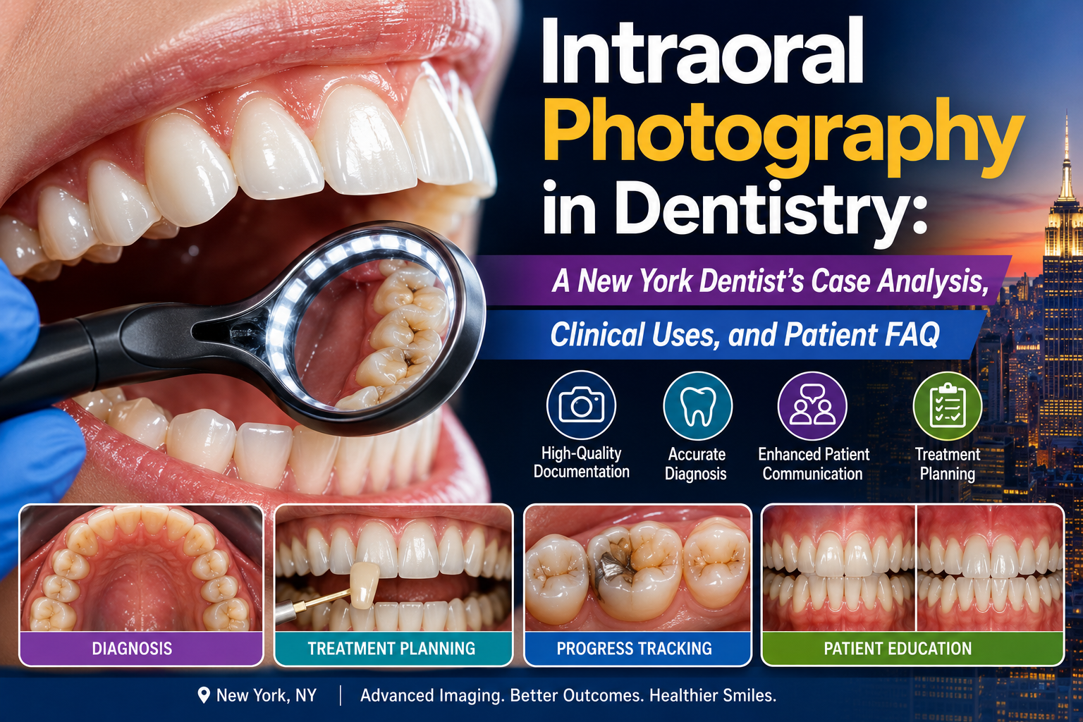
As a dentist practicing in New York, I use intraoral photography as an important visual component of a comprehensive dental examination.
An intraoral photograph is more than a picture of someone's teeth. When properly captured, it can document the appearance of the teeth, gums, restorations, oral tissues, and other clinical findings at a specific point in time.
The American Dental Association (ADA) recognizes photographs and intraoral photographs as components of dental records. Dental records can also include clinical notes, diagnostic charts, radiographs, treatment plans, and other diagnostic information.
For a patient, this means something very practical: your dentist can show you what they are seeing instead of relying entirely on verbal descriptions.
Educational disclaimer: This article presents a hypothetical U.S. dental case for educational purposes. It does not diagnose an individual patient or replace an examination by a licensed dentist.
What Is Intraoral Photography?
Intraoral photography is the process of taking detailed photographs inside the mouth.
Depending on the purpose of the examination, photographs may document:
-
Front teeth
-
Upper teeth
-
Lower teeth
-
Right side of the bite
-
Left side of the bite
-
Occlusal surfaces
-
Gums
-
Individual teeth
-
Dental restorations
-
Missing teeth
-
Tooth fractures
-
Discoloration
-
Tooth wear
-
Plaque and calculus
-
Oral soft tissues
A dentist may use a dedicated intraoral camera or a professional dental camera with appropriate mirrors, retractors, lighting, and accessories.
The objective is not simply to obtain an attractive photograph.
The objective is to obtain a clinically useful image.
Why Do Dentists Take Intraoral Photographs?
There are several important reasons.
1. Documentation
A photograph can document what a tooth or oral tissue looked like at a particular appointment.
This can be especially useful when monitoring:
-
Tooth fractures
-
Tooth wear
-
Gingival recession
-
Gingival inflammation
-
Discoloration
-
Existing restorations
-
Oral lesions
-
Cosmetic concerns
The ADA specifically lists photographs and intraoral photographs among information commonly maintained in dental records.
2. Patient Education
One of the most powerful applications is patient communication.
A dentist can point to a photograph and say:
"This is the area I'm concerned about."
The patient can then see the finding directly.
For example, instead of simply saying:
"You have significant wear on your lower front teeth,"
I can show the patient the actual tooth surfaces.
That can make treatment discussions much easier to understand.
3. Treatment Planning
Intraoral photographs can contribute to treatment planning alongside:
-
Clinical examination
-
Periodontal charting
-
Dental radiographs
-
Digital scans
-
Models
-
Patient history
-
Bite analysis
No single image should be treated as the entire diagnosis.
Instead, photographs become one piece of the clinical evidence.
The ADA has emphasized that diagnostic images—including clinical photographs—are routinely used by dentists to assess oral structures and support treatment planning.
4. Monitoring Changes Over Time
This is one of my favorite uses of dental photography.
Imagine photographing a patient's anterior teeth today and then comparing the images with photographs taken one year later.
We may be able to observe changes such as:
-
Increasing tooth wear
-
Gingival recession
-
Changes in inflammation
-
Restoration deterioration
-
Chipping
-
Tooth movement
-
Changes in oral hygiene
A photograph provides a visual baseline.
Case Analysis: A New York Patient With a Chipped Front Tooth
Patient Profile
Location: New York, USA
Age: 43
Sex: Male
Chief complaint: "I noticed a small chip on my front tooth."
The patient reports that the chip appeared while eating.
He has no severe pain and no facial swelling.
During the examination, I notice a small irregularity on the edge of an upper central incisor.
Rather than simply documenting:
"Small chip on anterior tooth."
I capture a properly framed intraoral photograph.
Step 1: Establish a Visual Baseline
The photograph shows:
-
Tooth #8 with a small incisal defect
-
Surrounding enamel appears intact
-
No obvious gingival swelling
-
No visible large fracture
-
Adjacent tooth appears clinically intact
This photograph becomes part of the diagnostic documentation.
The ADA notes that the dental record should contain accurate documentation of diagnostic information and treatment, and specifically includes photographs and intraoral photographs among examples of record content.
Step 2: Photograph From Multiple Angles
One photograph may not provide enough information.
I may capture:
Frontal view
Shows the overall appearance of the anterior teeth.
Right lateral view
Shows the relationship between the upper and lower teeth.
Left lateral view
Provides the opposite side.
Occlusal view
Helps evaluate the biting surfaces.
Close-up
Allows examination of the chipped area.
The exact views should be selected according to the clinical question.
Step 3: Combine Photography With Clinical Examination
This is critical.
A photograph cannot tell me everything.
For the chipped tooth, I would also evaluate:
-
Tooth mobility
-
Percussion
-
Sensitivity
-
Pulp status when indicated
-
Periodontal tissues
-
Occlusion
-
Possible crack extension
-
Adjacent teeth
-
History of trauma
If clinically indicated, radiographs may also be considered.
Current ADA recommendations emphasize that dental imaging should be selected based on the individual patient's history and clinical examination rather than using a one-size-fits-all approach.
Case Analysis: What Does the Photograph Tell Me?
Suppose the photograph reveals a small enamel fracture with no obvious extension below the gumline.
The patient has:
-
No spontaneous pain
-
No significant sensitivity
-
No mobility
-
No swelling
-
Normal surrounding soft tissues
-
Small localized enamel defect
This would be a very different situation from a patient whose photograph reveals a large fracture extending toward the gingival margin.
The image helps establish the visible extent of the problem.
But the photograph alone does not establish the complete diagnosis.
Another Case: Photographing Gingival Recession
Consider a 55-year-old patient who says:
"My front teeth look longer than they used to."
An intraoral photograph shows noticeable recession around several lower anterior teeth.
The photograph documents the appearance.
But I would also perform a periodontal evaluation.
That could include:
-
Probing depths
-
Clinical attachment levels
-
Bleeding assessment
-
Gingival measurements
-
Plaque assessment
-
Tooth mobility
-
Occlusal evaluation
The photograph gives me the visual evidence.
The periodontal examination gives me quantitative clinical information.
Together, they provide a much stronger assessment.
Intraoral Photography and Periodontal Disease
Photographs can be particularly useful for documenting visible periodontal findings.
For example:
Initial visit
Red, swollen gingival margins.
↓
After periodontal treatment
Reduced inflammation.
↓
Maintenance visit
More stable gingival appearance.
The images can help patients understand the visible changes associated with treatment.
However, photographs should not replace periodontal probing or other appropriate diagnostic procedures.
Intraoral Photography and Cosmetic Dentistry
Photography is extremely useful in cosmetic treatment planning.
For patients considering:
-
Porcelain veneers
-
Dental bonding
-
Crowns
-
Tooth whitening
-
Smile design
-
Orthodontics
-
Composite restorations
photographs can document:
-
Tooth shape
-
Tooth proportions
-
Shade
-
Gum contours
-
Incisal edges
-
Smile symmetry
-
Existing restorations
-
Tooth wear
Before-and-after photographs can also help communicate treatment changes.
However, photographs should be captured consistently if they are intended for meaningful comparison.
Intraoral Photography Before Veneers
Consider a patient requesting porcelain veneers for the upper front six teeth.
Before recommending treatment, I would want to evaluate:
-
Enamel availability
-
Existing restorations
-
Tooth position
-
Periodontal health
-
Occlusion
-
Tooth wear
-
Functional habits
-
Shade
-
Smile line
-
Gingival contours
Photographs can document the starting condition.
This is especially important because veneers are an irreversible or minimally reversible treatment depending on the preparation approach.
A photograph cannot determine whether veneers are appropriate by itself.
Intraoral Photography Before Dental Implants
Photography can also be useful during implant treatment planning.
For a missing tooth, an intraoral photograph can document:
-
Existing tooth loss
-
Gingival architecture
-
Adjacent teeth
-
Soft-tissue appearance
-
Prosthetic space
-
Existing restorations
However, implant planning also requires appropriate clinical evaluation and, when indicated, radiographic or 3D imaging.
The photograph provides the visible clinical perspective.
Radiographic imaging provides information that cannot be obtained from a standard photograph.
Photography Versus X-Rays
Patients sometimes ask:
"If you took photographs, why do I need an X-ray?"
The answer is simple:
They show different things.
Intraoral photograph
Primarily shows visible surfaces and tissues.
Dental radiograph
Can provide information about structures that are not directly visible, such as:
-
Tooth roots
-
Bone
-
Interproximal areas
-
Periapical structures
The ADA's 2026 recommendations emphasize that radiographs should be selected according to the patient's individual clinical needs after examination.
So photographs and radiographs should not be viewed as competing technologies.
They answer different clinical questions.
How a Dentist Captures a High-Quality Intraoral Photograph
A clinically useful photograph requires more than simply pointing a camera into the mouth.
Important factors include:
Patient positioning
The patient's head and mouth should be positioned consistently.
Retraction
Cheek and lip retractors help expose the teeth and gingiva.
Moisture control
Saliva can create reflections and obscure important surfaces.
Lighting
Adequate illumination is essential.
Focus
The area of clinical interest should be sharp.
Magnification
The image should provide enough detail without unnecessary background.
Orientation
Images should be captured consistently so that comparisons are meaningful.
Identification
The photograph should be correctly associated with the patient and clinical record.
The ADA states that diagnostic images should be correctly identified and maintained as part of the clinical record, with confidentiality protected under applicable privacy requirements.
Why Standardization Matters
Suppose I photograph a patient's teeth today from 30 cm away and six months later from 60 cm away.
Comparing the two photographs becomes much more difficult.
For meaningful monitoring, dental practices should try to maintain consistency in:
-
Camera position
-
Magnification
-
Lighting
-
Retraction
-
Angulation
-
Background
-
Patient positioning
Standardization makes changes easier to identify.
Intraoral Camera vs Professional Dental Photography
Intraoral Camera
Advantages include:
-
Quick
-
Convenient
-
Excellent for individual teeth
-
Useful for patient education
-
Easy integration into many dental records
Professional Dental Camera
Advantages can include:
-
Greater control over lighting
-
Higher image quality
-
More standardized documentation
-
Better full-mouth photography
-
Useful for cosmetic and restorative cases
The appropriate system depends on the clinical purpose and practice workflow.
Case Analysis: Detecting a Problem the Patient Could Not See
A 38-year-old New York patient comes for a routine examination.
She has no pain.
She says:
"I don't think anything is wrong with my teeth."
During intraoral photography, the dentist captures a close-up of a posterior tooth.
The photograph shows an area of discoloration and a questionable surface defect.
The dentist does not diagnose the condition from the photograph alone.
Instead, the finding triggers a closer clinical examination.
This is an important concept:
Photography can help identify something that deserves further investigation.
It does not mean every visible discoloration represents disease.
Intraoral Photography and Patient Communication
One of the biggest advantages is improving the dentist-patient conversation.
Imagine showing a patient:
Photograph → Clinical finding → Treatment options
Instead of giving the patient a purely verbal explanation, the dentist can visually demonstrate:
"This is the fractured area."
"This is where the gum has receded."
"This restoration has a visible defect."
"This is the area we want to monitor."
This can improve understanding and help patients participate in treatment decisions.
Photography as Part of the Dental Record
Intraoral photographs are clinical records.
That means practices should treat them with the same professionalism applied to other patient information.
The ADA explains that dental records are official clinical documents and that accurate documentation is important for patient care and professional risk management.
Photographs should therefore be:
-
Correctly associated with the patient
-
Stored securely
-
Clinically relevant
-
Properly documented
-
Protected as confidential health information
Patients also have rights regarding access to copies of their dental records, subject to applicable federal and state requirements.
Intraoral Photography and Dental Insurance
Photographs may also become relevant when documenting clinical findings for treatment planning or insurance-related processes.
However, a photograph should not be created merely because an insurer requests an image if there is no appropriate clinical reason.
The dentist remains responsible for accurate clinical documentation and appropriate treatment decisions.
The ADA emphasizes that documentation should accurately reflect the patient's findings, diagnosis, treatment, and treatment planning.
Intraoral Photography and AI
Dental imaging is increasingly becoming connected with digital workflows and artificial intelligence.
The ADA is developing standards related to dental image analysis and AI, including standards addressing validation datasets for image-analysis systems.
This creates exciting possibilities for:
-
Image organization
-
Detection assistance
-
Documentation
-
Comparison of images
-
Clinical decision support
But AI should not replace the dentist's clinical examination.
A photograph is an input.
The dentist remains responsible for interpreting the patient's overall clinical condition.
Frequently Asked Questions
What is intraoral photography?
It is the use of photography to document visible structures inside the mouth, including teeth, gums, restorations, and oral tissues.
Why does my dentist take pictures of my teeth?
Photographs can help document clinical findings, educate you about your oral condition, monitor changes, and support treatment planning.
Can an intraoral photograph diagnose a cavity?
A photograph may reveal a suspicious area, but the dentist may need additional clinical examination and sometimes radiographic evaluation before establishing a diagnosis.
Can a dentist see cavities in a photograph?
Some visible cavities or defects may be apparent, but many dental problems cannot be reliably identified from a photograph alone.
Are dental photographs part of my medical record?
Intraoral photographs are commonly maintained as part of the dental record. The ADA specifically includes intraoral photographs among examples of information contained in dental records.
Can I request copies of my dental photographs?
Patients generally have rights to access and obtain copies of their dental records under applicable federal and state requirements. Practices should follow HIPAA and relevant state requirements when releasing records.
Are my dental photographs private?
They should be handled as confidential patient information. Dental practices must follow applicable privacy and security requirements, including HIPAA where applicable.
Do I need X-rays if my dentist takes photographs?
Sometimes. Photographs and radiographs provide different information. X-rays are selected when clinically necessary to answer diagnostic or treatment-planning questions.
Can intraoral photographs show gum disease?
They can show visible signs such as redness, swelling, recession, or other changes, but periodontal diagnosis may require probing and other clinical measurements.
Can photographs show tooth wear?
Yes. Photography can be very useful for documenting tooth wear and comparing its appearance over time.
Are intraoral photographs useful before veneers?
Yes. They can document the starting condition and help evaluate tooth shape, color, gum contours, restorations, and smile characteristics. However, they are only one component of treatment planning.
Are intraoral photographs useful for dental implants?
Yes. They can document the visible condition of the missing-tooth area and surrounding teeth and soft tissues. Implant planning may also require clinical examination and appropriate imaging.
Can my dentist use an old photograph to diagnose my current condition?
Not by itself. A photograph represents the patient's condition at the time it was taken. Current diagnosis requires current clinical assessment when appropriate.
New York Case Analysis: The Value of a Baseline Photograph
Let's return to our hypothetical New York patient with the chipped front tooth.
Visit 1
Photograph documents a small enamel fracture.
Visit 2
The tooth remains stable.
Visit 3
A second photograph shows a new area of wear.
Now the dentist has something extremely valuable:
A visual timeline.
Instead of relying entirely on memory, the clinician can compare documented images.
This can help answer:
-
Is the fracture changing?
-
Is the wear progressing?
-
Is the restoration stable?
-
Has gingival recession changed?
-
Is the patient maintaining oral hygiene?
-
Does treatment need to be reconsidered?
The Dentist's Clinical Perspective
From my perspective as a New York dentist, one of the biggest advantages of intraoral photography is that it turns an invisible clinical conversation into something the patient can actually see.
A patient may not understand what a:
fractured cusp
or
gingival recession
or
worn incisal edge
looks like.
But when the photograph is displayed on a monitor, the conversation changes.
The patient can participate in the examination.
That is powerful.
However, I would always emphasize one rule:
A photograph is evidence—not the entire diagnosis.
The best dental decisions combine:
Patient history
Clinical examination
Intraoral photography
Periodontal assessment when indicated
Radiographs or other imaging when clinically justified
Occlusal and functional evaluation when appropriate
That complete picture provides a much stronger foundation for diagnosis and treatment planning.
Final Takeaway
Intraoral photography has become an important part of modern digital dentistry.
For patients, it can provide:
-
Better understanding
-
Better communication
-
Visual documentation
-
Treatment monitoring
-
Improved treatment discussions
For dentists, it can provide:
-
A clinical baseline
-
Documentation of visible findings
-
Support for treatment planning
-
Before-and-after comparison
-
Better patient education
The American Dental Association recognizes intraoral photographs as part of dental records, and current dental imaging guidance continues to emphasize individualized clinical assessment and appropriate use of diagnostic images.
The most important principle is simple:
Take the photograph for a clinical purpose, document it accurately, protect the patient's privacy, and interpret it together with the complete dental examination.
A well-captured intraoral photograph does not replace the dentist.
It helps the dentist—and the patient—see the dentistry more clearly.
Related Clinical Cases
Patients with lower front gum inflammation may also experience tartar buildup, periodontal disease, gum abscesses, or tooth decay. Explore these related clinical case studies.


Other topics:
Other Dental Clinic