Ear Pain After Tooth Extraction: Causes, Relief & When to Worry
Severity:
Teeth Problems:
A Clinical Guide from an Idaho Dental Bone Graft Expert (10 Years Experience)
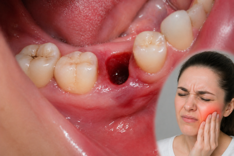
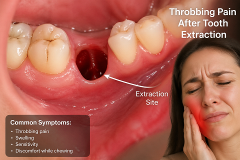
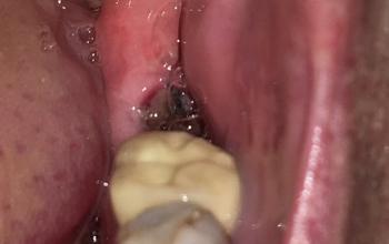
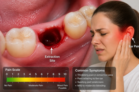
Ear pain after a tooth extraction can feel alarming. Many patients assume something is wrong with their ear—but in most cases, the source is actually your dental procedure.
After 10 years of treating extraction and implant cases in Idaho, I can tell you this clearly:
Ear pain after tooth extraction is usually referred pain—and often completely normal.
But timing and pattern matter. In this guide, you’ll learn: