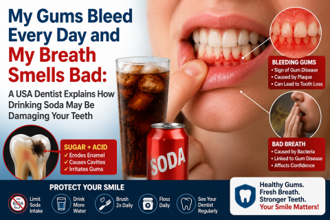
My Gums Bleed Every Day and My Breath Smells Bad: A USA Dentist Explains How Drinking Soda May Be Damaging Your Teeth
Topics:
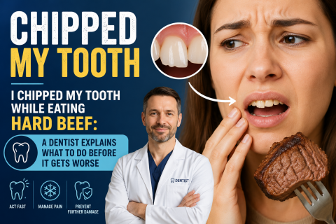
"I was just enjoying my meal. The beef was tougher than I expected. As I chewed down, I suddenly heard a small crunch. At first I thought it was part of the meat. Then I realized a tiny piece of my own tooth had broken off.
Now I'm anxious.
Did I permanently damage my tooth?
Will I lose it?
Will it become infected?
Should I rush to the dentist today?"
If this sounds familiar, you are not alone.
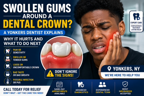
"I've been worrying all night. My gums are swollen around my crown, the pain keeps getting worse, my head feels like it's spinning, and I can't sleep. I've tried brushing, rinsing, and taking pain medication, but nothing seems to help."
If this sounds familiar, you're not alone.
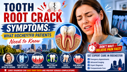
"I've been calling dental offices all morning. The pain is getting worse. My tooth hurts every time I bite down. My gum is swollen, and nobody seems to be answering quickly enough."
If this sounds familiar, you are not alone.
Many patients experiencing a fractured tooth root feel frustrated, anxious, and even frightened when they cannot immediately reach a dentist. Dental pain can become overwhelming, especially when it interferes with eating, sleeping, working, or simply getting through the day.
A dental emergency can strike without warning. One moment you are enjoying your day, and the next you are dealing with severe tooth pain, facial swelling, a broken tooth, or a dental infection that makes it difficult to eat, sleep, or even concentrate. When pain becomes unbearable, many patients begin searching online for answers, especially regarding how much an emergency tooth extraction will cost and how quickly treatment can be provided.
 |
 |
 |
 |
 |
 |

![]()