Where Bright Smiles Begin.

Bone Loss and Facial Bone Loss: A Swiss Dental Expert’s Clinical Review, Case Analysis, and Patient Advice
image hero:
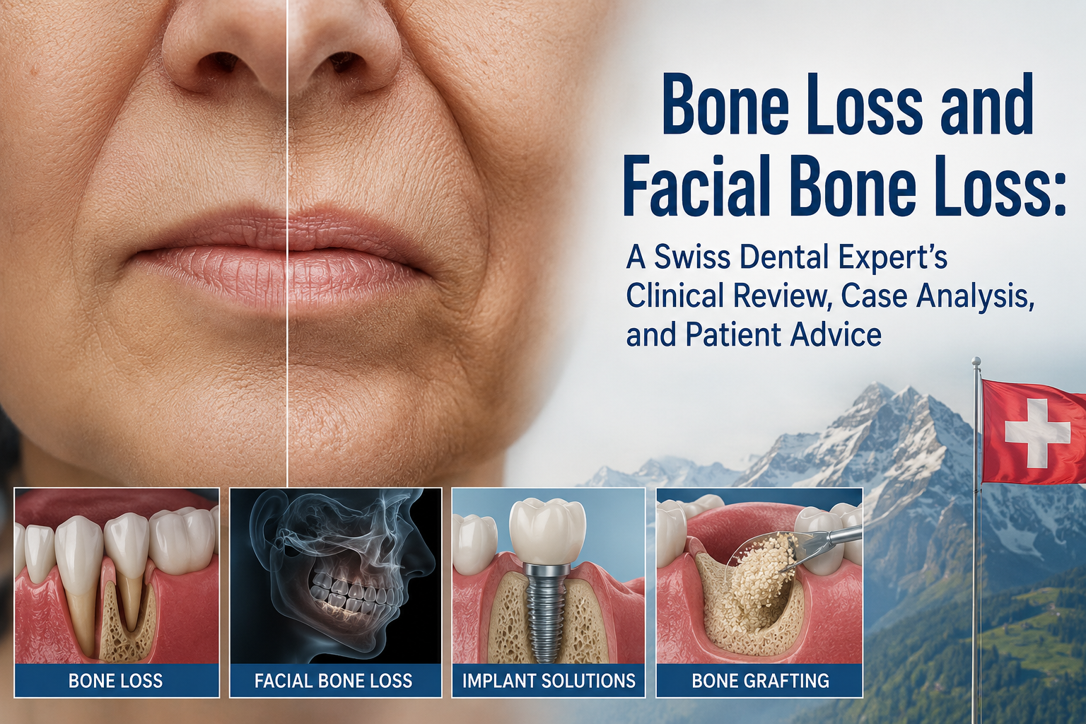
Facial bone loss is more than a cosmetic problem. In dentistry, loss of the alveolar bone surrounding the teeth can progressively change the shape of the jaw, gums, lips, cheeks, and overall facial profile.
A patient may notice a missing tooth, gum recession, a sunken area around the mouth, or changes in the way the teeth meet. However, the underlying problem can be occurring beneath the gum line: the supporting bone may be gradually disappearing.
The key clinical question is not simply, “How much bone has been lost?” It is:
Why was the bone lost, how much supporting structure remains, and can the remaining anatomy be stabilized or reconstructed?
The American Academy of Periodontology notes that tooth extraction is followed by dimensional reduction of the alveolar process, while severe ridge atrophy can significantly complicate later implant treatment.
1. What Is Facial Bone Loss?
The term facial bone loss can refer to several different anatomical problems.
In dentistry, the most important is usually alveolar bone loss—loss of the bone that surrounds and supports the teeth.
This is different from generalized loss of the facial skeleton caused by trauma, developmental conditions, tumors, or other medical disorders.
When a tooth is lost, the surrounding alveolar bone no longer receives the same functional stimulation from the tooth root. Over time, the ridge can become narrower and/or shorter.
This may produce:
- A depression in the gum
- A narrow dental ridge
- Gum recession
- Reduced support for neighboring teeth
- Changes in facial soft-tissue support
- A sunken appearance around a missing tooth
- Difficulty placing a conventional dental implant
- Changes in chewing and bite relationships
The extent of the visible facial change depends on the location and amount of bone loss.
2. Major Causes of Dental and Facial Bone Loss
Periodontal Disease
Advanced periodontal disease is one of the major causes of tooth-supporting bone destruction.
Inflammation associated with periodontal disease can progressively damage the tissues supporting a tooth.
As the supporting bone decreases, the tooth may become increasingly mobile.
Clinical warning signs include:
- Bleeding gums
- Persistent gum inflammation
- Gum recession
- Bad breath
- Increasing tooth mobility
- Teeth appearing longer
- New spaces developing between teeth
- Changes in the bite
The important point is that periodontal bone loss is not simply an aesthetic problem. It can ultimately compromise the stability of the natural dentition.
Tooth Loss
Tooth loss itself can initiate a remodeling process in the alveolar ridge.
The bone that once surrounded the tooth root can progressively resorb after extraction.
This is especially important when the patient plans to replace the missing tooth with an implant later.
The AAP specifically describes how absence of a tooth can produce dimensional reduction of the alveolar process and potentially create challenges for future implant-supported rehabilitation.
Swiss Expert Review
From an implant-planning perspective, waiting several years after losing a tooth and then asking for an implant is not always the simplest pathway.
A patient should ideally have the extraction site evaluated early, particularly when implant replacement is being considered.
3. Bone Loss Can Change the Face
The jawbone provides structural support for the teeth and surrounding soft tissues.
When substantial bone and tooth support are lost, the lips, cheeks, and gingival architecture may also change.
A patient with extensive tooth loss may gradually develop:
- Reduced lower-face support
- Increased appearance of facial wrinkles around the mouth
- A more collapsed oral profile
- Reduced lip support
- Narrowing of the dental ridge
- Changes in facial symmetry
However, facial aging should not automatically be attributed to dental bone loss.
A proper diagnosis must distinguish normal age-related skeletal and soft-tissue changes from localized or generalized dental bone loss.
4. Clinical Case Analysis
Case Example: Long-Term Missing Upper Front Tooth
Consider a patient who lost an upper front tooth several years ago.
Initially, the patient may have replaced the tooth with a temporary or removable prosthesis.
Years later, the patient notices a depression in the gum and a slight hollow appearance above the missing tooth.
The patient now requests a dental implant.
Clinical Interpretation
The problem is no longer simply:
“There is a missing tooth.”
The problem may now involve:
Missing tooth + reduced ridge width + reduced ridge height + soft-tissue deficiency + altered esthetics.
A clinical examination would therefore need to evaluate:
- Remaining bone volume
- Ridge width
- Ridge height
- Adjacent tooth roots
- Periodontal health
- Soft-tissue thickness
- Occlusion
- Available implant position
- Sinus anatomy when relevant
- Overall medical and dental risk factors
A three-dimensional CBCT examination may be considered when clinically indicated to evaluate the available anatomy for implant planning.
5. Why an Implant Cannot Simply Be “Screwed Into” Any Bone
Dental implants require adequate supporting anatomy.
The AAP explains that successful implant treatment depends on sufficient bone quantity and quality, together with healthy periodontal tissues.
If the ridge has become severely resorbed, placing an implant directly into the deficient area may compromise:
- Implant positioning
- Esthetics
- Stability
- Prosthetic design
- Long-term tissue maintenance
This is why some patients require bone augmentation before or during implant placement.
6. Bone Grafting and Ridge Reconstruction
Bone augmentation is designed to rebuild deficient areas of the jaw.
Depending on the defect, treatment may include:
Ridge Preservation
Performed around the time of extraction in selected cases to help maintain the dimensions of the extraction site.
Guided Bone Regeneration
A bone graft and membrane or other regenerative approach may be used to reconstruct a deficient ridge.
Ridge Augmentation
Used when the existing ridge lacks sufficient width and/or height for the planned restoration.
Sinus Augmentation
In the posterior upper jaw, loss of bone combined with the anatomy of the maxillary sinus can make implant placement challenging.
Sinus augmentation can create additional bone volume in selected patients. The AAP notes that bone development after sinus augmentation may require several months before implant placement, although treatment protocols vary and simultaneous placement can sometimes be appropriate.
Autogenous or Other Bone-Grafting Materials
The choice of graft material depends on:
- Defect size
- Defect morphology
- Implant plan
- Patient factors
- Clinician experience
- Required amount of augmentation
There is no single graft material that is automatically best for every patient.
7. Clinical Review: When Is Bone Grafting Particularly Important?
Bone grafting may be considered when there is insufficient ridge anatomy for the intended restoration.
The AAP describes ridge modification as a procedure that can rebuild areas where inadequate bone remains for implant placement.
However, the phrase “You need a bone graft” should never replace a complete diagnosis.
A responsible treatment plan should explain:
- Where the bone is deficient
- Whether the deficiency is horizontal, vertical, or both
- Why augmentation is necessary
- What grafting technique is proposed
- Whether soft-tissue augmentation is also required
- Whether the implant can be placed simultaneously
- Expected healing period
- Alternatives
- Risks
- Long-term maintenance
8. Bone Loss Around Dental Implants
A common misconception is that once an implant has been placed, bone loss is no longer a concern.
That is incorrect.
Dental implants can develop inflammatory disease around the implant.
The AAP distinguishes peri-implant mucositis from peri-implantitis. Peri-implantitis involves inflammation accompanied by deterioration of the bone supporting the implant.
Potential warning signs include:
- Bleeding around an implant
- Red or swollen tissue
- Suppuration
- Increasing discomfort
- Gum recession
- Exposed implant components
- Increasing probing depths
- Radiographic bone loss
- Changes in implant stability in advanced cases
Expert Advice
An implant should not be considered a “maintenance-free artificial tooth.”
It requires professional monitoring and meticulous home care.
9. Case Analysis: Severe Bone Loss Around an Implant
Imagine a patient who received an implant five years ago.
The patient now reports occasional bleeding when brushing.
The implant appears clinically stable, so the patient assumes there is no problem.
Radiographic examination, however, demonstrates progressive bone loss around the implant.
Clinical Interpretation
This could represent peri-implant disease rather than a simple superficial gum problem.
The clinician must evaluate:
- Plaque control
- Implant position
- Prosthetic design
- Occlusion
- Periodontal history
- Smoking status
- Systemic risk factors
- Radiographic bone changes
- Previous treatment
Treatment should be individualized according to the severity and cause of disease.
10. Can Facial Bone Loss Be Reversed?
This requires an important distinction.
Some bone defects can be reconstructed or regenerated, but not every type or degree of bone loss can be completely restored to its original anatomy.
Regenerative procedures can improve deficient areas in appropriately selected cases.
The AAP describes ridge augmentation and sinus augmentation as established approaches for developing additional bone where implant placement is otherwise compromised.
The biological potential for regeneration depends heavily on:
- Defect morphology
- Remaining bone
- Blood supply
- Soft-tissue condition
- Infection control
- Patient factors
- Surgical technique
- Healing
- Maintenance
Therefore, patients should be cautious of advertisements promising “100% bone regeneration” or guaranteed restoration of the original facial structure.
11. Prevention Is Often Better Than Reconstruction
From a Swiss evidence-oriented dental perspective, the best bone-regeneration procedure is often the one that can be avoided through early prevention and appropriate treatment.
Important preventive measures include:
Maintain Natural Teeth Whenever Possible
The AAP emphasizes preservation of natural teeth whenever they can be predictably maintained rather than unnecessarily extracting salvageable teeth.
Treat Periodontal Disease Early
Controlling periodontal inflammation can help prevent further destruction of supporting tissues.
Plan Extractions Carefully
When extraction is unavoidable and future implant treatment is anticipated, ridge-preservation strategies may be considered.
Replace Missing Teeth Appropriately
A missing tooth should be evaluated rather than simply ignored for many years.
Maintain Implants
Implants require brushing, interdental cleaning, professional monitoring, and long-term maintenance.
12. Patient Advice: When Should You Seek an Evaluation?
You should consider a comprehensive dental examination if you notice:
- A loose tooth
- Progressive gum recession
- Bleeding gums
- Persistent bad breath
- A missing tooth
- A visible depression in the jaw
- Facial asymmetry around a missing tooth
- Difficulty wearing a denture
- An implant that bleeds
- Gum swelling around an implant
- Changes in your bite
- Increasing tooth mobility
The earlier the underlying cause is identified, the more treatment options may remain available.
13. Questions to Ask Your Dentist or Periodontist
Before proceeding with extensive bone reconstruction or implant treatment, ask:
1. What caused my bone loss?
2. How much bone have I actually lost?
3. Is the loss horizontal, vertical, or both?
4. Can my natural tooth still be saved?
5. Do I need periodontal treatment before reconstruction?
6. Do I need a bone graft?
7. Do I need soft-tissue grafting as well?
8. Can the implant be placed immediately, or must the site heal first?
9. How long will reconstruction take?
10. What are the alternatives to an implant?
11. What are the risks and expected complications?
12. How will the result be maintained long term?
These questions help move the conversation away from simply choosing a procedure and toward proper diagnosis and treatment planning.
14. Dental Hub Review: What I Would Look for in a High-Quality Bone-Loss Treatment Plan
A strong treatment plan should demonstrate five things:
1. Diagnosis Before Treatment
The clinician should identify the cause of the bone loss.
2. Preservation Before Replacement
If a natural tooth can reasonably be preserved, that option should be evaluated.
3. Three-Dimensional Planning
Implant reconstruction should consider the available bone, soft tissues, prosthetic requirements, and facial esthetics.
4. Regeneration When Indicated
Bone grafting should be recommended because the anatomy requires it—not simply because it is an additional procedure.
5. Long-Term Maintenance
The treatment plan should include prevention of recurrent periodontal or peri-implant disease.
15. Swiss Expert Perspective
In Switzerland, where precision, preventive care, and long-term treatment planning are highly valued, I would approach facial bone loss with one fundamental principle:
Do not treat the missing bone before understanding why the bone disappeared.
Bone loss is a biological diagnosis, not merely a cosmetic defect.
A successful reconstruction should restore more than the ability to place an implant. The objective should be to create a stable biological environment, appropriate function, acceptable esthetics, and a maintenance strategy that protects the result for many years.
For patients with severe bone loss, multidisciplinary collaboration between a general dentist, periodontist, oral surgeon, and restorative or prosthodontic specialist may be appropriate.
16. Final Dental Hub Advice
Facial bone loss should not be ignored.
A missing tooth, periodontal disease, poorly maintained implant, trauma, or advanced ridge resorption can progressively alter the supporting architecture of the mouth.
But bone loss does not automatically mean that reconstruction is impossible.
Modern periodontal and implant dentistry offers several approaches, including ridge preservation, guided bone regeneration, ridge augmentation, and sinus augmentation for appropriately selected cases.
The most important step is therefore not immediately choosing a bone graft or implant.
It is obtaining an accurate diagnosis and understanding the complete treatment sequence.
Preserve what can be saved. Control disease. Rebuild deficient anatomy when necessary. Restore function and esthetics. Then maintain the result.
This article is educational and does not replace an examination, periodontal evaluation, radiographs/CBCT when indicated, or individualized treatment planning by a qualified dental professional.
Related Clinical Cases
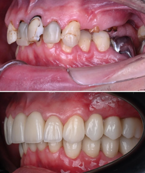
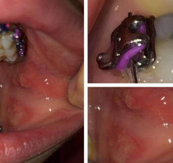
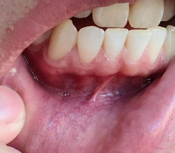
Patients with lower front gum inflammation may also experience tartar buildup, periodontal disease, gum abscesses, or tooth decay. Explore these related clinical case studies.


Other topics:
Other Dental Clinic