Strong Teeth, Fresh Breath, Brighter You.

Cone Beam Computed Tomography (CBCT): USA Technology & Clinical Review
image hero:
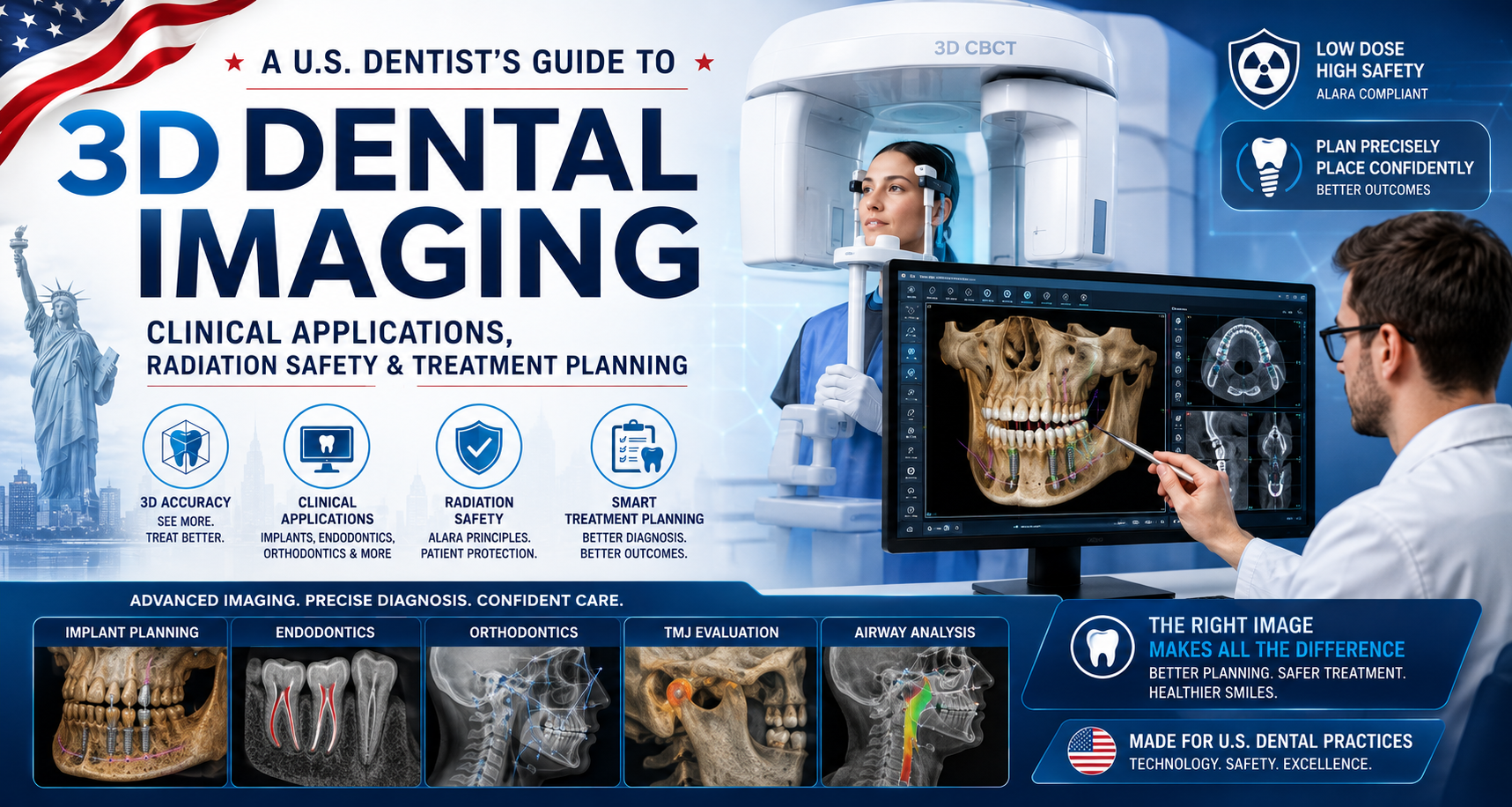
A U.S. Dentist’s Guide to 3D Dental Imaging, Clinical Applications, Radiation Safety and Treatment Planning
Cone Beam Computed Tomography (CBCT) has become one of the most important advanced imaging technologies in modern American dentistry. Unlike conventional intraoral X-rays and panoramic radiographs, which produce two-dimensional images, CBCT creates a three-dimensional representation of teeth and surrounding maxillofacial structures.
From an American dentist's perspective, however, the most important point is not simply that CBCT can show more.
The important question is:
Will the additional 3D information change the diagnosis, treatment plan or clinical management of this patient?
Current U.S. recommendations emphasize patient-specific, clinically justified imaging rather than routine CBCT scanning. The ADA's updated 2026 recommendations specifically address both 2D and 3D dental imaging and emphasize clinical examination and individualized patient selection.
What Is Dental CBCT?
CBCT is an X-ray-based imaging system that rotates around the patient's head while acquiring multiple projection images. Specialized software reconstructs these images into a three-dimensional volume composed of small units called voxels.
This allows the dentist or dental specialist to examine anatomy in multiple planes, including:
- Axial
- Sagittal
- Coronal
- 3D reconstructed views
The technology is particularly valuable for evaluating teeth, roots, alveolar bone, jaws and other maxillofacial structures.
The American Association of Endodontists describes CBCT as providing three-dimensional views that can significantly improve diagnosis and treatment planning in selected cases.
Why 3D Imaging Matters in Dentistry
A conventional periapical X-ray compresses three-dimensional anatomy into a flat image.
That can make structures overlap.
For example, two roots may appear superimposed on a 2D radiograph even though they are positioned differently in three-dimensional space.
CBCT allows the clinician to rotate through the volume and examine the anatomy from different directions.
This can be particularly valuable when the dentist needs to determine:
Where exactly is the structure?
How much bone is available?
Where is the nerve canal?
Is there an additional root canal?
Is there a fracture or resorption pattern that cannot be confidently identified on 2D imaging?
Major Clinical Applications in U.S. Dentistry
1. Dental Implant Planning
CBCT has become an important planning tool for implant dentistry.
Before implant placement, the clinician may need to evaluate:
- Available bone height
- Buccolingual bone width
- Ridge anatomy
- Adjacent roots
- Inferior alveolar canal
- Mental foramen
- Maxillary sinus
- Nasal cavity
- Other anatomical structures
This information can help the implant team determine the appropriate implant position and whether additional procedures, such as bone augmentation or sinus-related treatment, may be necessary.
CBCT can also be integrated with digital implant planning, surgical guides and navigation technologies.
2. Endodontics and Root Canal Treatment
CBCT can be particularly valuable in complex endodontic cases.
The technology may help clinicians evaluate:
- Additional canals
- Complex root morphology
- Calcified canals
- Root resorption
- Periapical pathology
- Root fractures
- Previously treated teeth
- Failed root canal treatment
- Surgical anatomy
One well-known example is the MB2 canal in maxillary molars, which can sometimes be difficult to identify using conventional 2D radiography.
The AAE/AAOMR position emphasizes that CBCT should be used selectively rather than routinely for endodontic diagnosis. Their updated 2025 guidance continues to emphasize individualized indications and radiation optimization.
3. Impacted Wisdom Teeth and Complex Extractions
CBCT can provide valuable information before certain surgical extractions.
For example, an impacted mandibular third molar may have a close relationship with the inferior alveolar canal.
A two-dimensional panoramic image may suggest a potentially close relationship, but CBCT can provide additional information about the three-dimensional relationship.
This can help the oral surgeon evaluate surgical risk and determine the most appropriate treatment strategy.
CBCT does not eliminate surgical complications, but better anatomical information can improve planning.
4. Orthodontics
CBCT may be used selectively in orthodontic cases involving:
- Impacted teeth
- Supernumerary teeth
- Complex tooth positioning
- Skeletal abnormalities
- Craniofacial anatomy
- Certain surgical orthodontic procedures
However, CBCT should not automatically replace conventional orthodontic imaging.
The scan should provide information that is clinically useful enough to justify the additional radiation exposure.
5. Airway and Maxillofacial Assessment
Three-dimensional imaging can provide information about airway anatomy and surrounding skeletal structures.
CBCT may be incorporated into certain orthodontic, oral-surgical and maxillofacial evaluations.
However, an important clinical distinction is necessary:
CBCT airway measurements should not automatically be interpreted as a diagnosis of obstructive sleep apnea.
Sleep apnea diagnosis requires appropriate clinical evaluation and, when indicated, formal sleep testing.
CBCT vs. 2D Dental X-Ray vs. Medical CT
| Feature | 2D Dental X-Ray | Dental CBCT | Medical CT |
|---|---|---|---|
| Image | 2D | 3D | 3D |
| Primary strength | Routine dental diagnosis | High-resolution dental/maxillofacial anatomy | Broader medical imaging |
| Hard-tissue detail | Good | Excellent for many dental applications | Excellent |
| Soft-tissue evaluation | Limited | Limited | Generally superior |
| Typical setting | Dental office | Dental/specialty office or imaging center | Hospital/imaging center |
| Radiation | Generally lowest | Variable; depends heavily on protocol | Generally higher |
| Field of view | Limited | Selectable | Usually broader |
| Main advantage | Simple and efficient | 3D anatomical information | Comprehensive medical evaluation |
One important correction to many simplified CBCT comparisons: there is no single universal CBCT radiation dose. Dose varies substantially according to machine, field of view, voxel size, exposure settings and clinical protocol.
Therefore, quoting one fixed number for "CBCT radiation" can be misleading.
Radiation Safety: The U.S. Approach
From a U.S. dental expert perspective, CBCT should never be ordered simply because the technology is available.
The fundamental principle is:
Justification + Optimization
The ADA describes the ALARA principle — As Low As Reasonably Achievable — as an important component of dental radiographic safety. The clinician should ensure that an X-ray examination has a clinical benefit and that necessary exposure is minimized.
The newer terminology ALADAIP — As Low As Diagnostically Acceptable, Indication-oriented and Patient-specific — is also emphasized in the updated AAE/AAOMR endodontic guidance.
Field of View: One of the Most Important CBCT Decisions
Modern CBCT systems can often use different fields of view (FOVs).
A limited FOV may focus on:
- One tooth
- Several teeth
- One implant site
- A specific region of the jaw
A larger FOV can capture:
- An entire arch
- Both jaws
- Broader craniofacial anatomy
When the diagnostic question involves a small region, a smaller field of view may reduce unnecessary exposure while providing the information needed.
This is one reason modern CBCT protocols should be indication-specific rather than one-size-fits-all.
CBCT for Children: Extra Caution
Children and adolescents require particular attention when ionizing radiation is used.
The goal is not to avoid medically necessary imaging.
The goal is to ensure that imaging is clinically justified and appropriately optimized for the individual patient.
The ADA notes that dental imaging should be selected based on factors such as age, oral-health status, disease risk, symptoms and clinical findings. The organization also supports child-specific radiation optimization initiatives.
For a child, a dentist should therefore ask:
Can the clinical question be answered without CBCT?
If not:
What is the smallest appropriate field of view and lowest suitable exposure that provides adequate diagnostic information?
CBCT Is Not a Substitute for a Clinical Examination
This is one of the most important concepts in modern dental imaging.
A CBCT scan is a diagnostic adjunct.
It does not replace:
- Patient history
- Clinical examination
- Periodontal assessment
- Pulp testing
- Percussion
- Palpation
- Conventional radiographs when indicated
- Specialist evaluation
The ADA's current recommendations emphasize performing a clinical examination before imaging and determining whether imaging is necessary for diagnosis, treatment planning or clinical management.
The Pros of CBCT
1. Three-Dimensional Anatomy
The clinician can evaluate anatomy in three dimensions rather than relying exclusively on a flat image.
2. Excellent Dental Hard-Tissue Visualization
CBCT is particularly useful for teeth and osseous structures.
3. Treatment Planning
Implant placement, complex extractions and selected endodontic procedures can benefit from 3D anatomical information.
4. Detection of Complex Anatomy
Additional canals, unusual root morphology and certain pathological findings may become easier to evaluate.
5. Digital Integration
CBCT datasets can be integrated with:
- Digital impressions
- Implant-planning software
- CAD/CAM workflows
- Surgical guides
- Dynamic navigation
- Some AI-assisted imaging workflows
The AAE has specifically highlighted CBCT's role in technologies such as 3D printing, surgical guides and dynamic navigation.
Limitations and Risks
CBCT is powerful, but it is not perfect.
Radiation Exposure
CBCT generally involves more radiation than a single routine intraoral radiograph, although actual dose varies substantially between examinations.
Metal Artifacts
Crowns, fillings, implants and other metallic objects can produce artifacts that interfere with image interpretation.
Limited Soft-Tissue Contrast
CBCT is primarily optimized for hard-tissue and dental/maxillofacial applications. It should not be considered equivalent to MRI for soft-tissue imaging.
Incidental Findings
A large scan can reveal findings outside the original clinical question.
This creates another responsibility:
Someone appropriately trained must review the relevant imaging volume.
The more information a scan provides, the greater the importance of competent interpretation.
An Important 2026 Safety Update: Patient Shielding
Older dental information frequently tells patients that lead aprons and thyroid collars should routinely be used for dental X-rays.
Current U.S. recommendations have changed.
The ADA's 2024 radiation-safety recommendations no longer recommend routine use of lead abdominal aprons or thyroid collars for dental radiography, because shielding can interfere with the X-ray beam and potentially lead to repeat imaging. Proper positioning, collimation and dose optimization are emphasized instead.
However, state-specific laws and regulations can differ, so dental offices must follow applicable requirements.
Does Every Dental Patient Need CBCT?
No.
This is perhaps the most important message of the entire CBCT review.
CBCT is not simply a "better X-ray."
It is a more advanced imaging modality that should be selected when its additional information provides sufficient clinical value.
A routine cavity examination may be adequately evaluated with clinical examination and appropriate 2D imaging.
A complex implant case may benefit substantially from CBCT.
A complicated retreatment case may benefit from a limited-FOV scan.
A complex impacted tooth may require three-dimensional anatomical assessment.
The decision should be individualized.
The AAE/AAOMR specifically states that CBCT should not be used routinely for endodontic screening in the absence of appropriate clinical indications.
What Does CBCT Cost in the USA?
Patient pricing varies considerably by:
- Geographic location
- Dental practice
- Imaging center
- Field of view
- Type of examination
- Insurance
- Whether the scan is included in a larger treatment package
A commonly quoted U.S. patient-pay range is approximately $150–$500, but this should be treated as a general market estimate rather than a national standard.
Insurance coverage also varies.
Some dental plans may classify CBCT as a diagnostic procedure, while coverage may depend on the clinical indication, policy exclusions and preauthorization requirements.
Patients should ask the dental office:
"Is the CBCT included in my treatment fee, or is it billed separately?"
and
"Will my dental or medical insurance cover this examination?"
The U.S. Dentist's Clinical Decision
When I evaluate whether a patient needs CBCT, I would frame the decision around one fundamental question:
What clinical question am I trying to answer?
If conventional imaging already provides the necessary information, CBCT may add unnecessary radiation without enough additional diagnostic benefit.
If conventional imaging is inconclusive and the 3D information could significantly change treatment, CBCT may be extremely valuable.
This is the essence of modern evidence-based dental imaging.
Final Clinical Review
CBCT represents a major technological advancement in American dentistry.
Its greatest advantage is not simply producing a beautiful 3D image.
Its real value is giving the clinician additional anatomical information that can influence diagnosis, treatment planning and clinical decision-making.
For implant dentistry, complex endodontics, impacted teeth and selected orthodontic or oral-surgical cases, CBCT can provide information that conventional 2D imaging cannot adequately demonstrate.
But more imaging does not automatically mean better dentistry.
The best CBCT examination is the one that is clinically justified, appropriately limited, properly acquired and correctly interpreted.
That is the modern U.S. approach:
Examine first.
Define the clinical question.
Select the appropriate imaging modality.
Use the smallest appropriate field of view.
Optimize radiation exposure.
Interpret the complete examination appropriately.
And most importantly:
Use 3D imaging when seeing in three dimensions can genuinely improve patient care.
Educational content only. CBCT selection, interpretation and treatment decisions should be made by a qualified dental professional based on the patient's individual clinical circumstances and applicable U.S. regulations.
Related Clinical Cases
Patients with lower front gum inflammation may also experience tartar buildup, periodontal disease, gum abscesses, or tooth decay. Explore these related clinical case studies.


Other topics:
Other Dental Clinic