Your Smile, Perfected with Precision.
Rampant Early Childhood Caries: Diagnosis, Urgent Management & Definitive Treatment Options
Image:
Severity:
Teeth Problems:
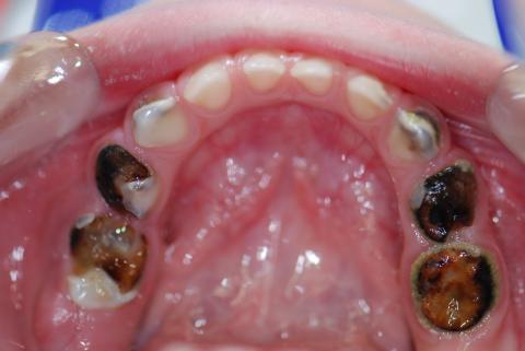
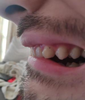
Immediate visual diagnosis (short)
This image shows severe, rampant early childhood caries (S-ECC) of the maxillary primary teeth with extensive coronal destruction on both sides. Multiple primary molars appear non-restorable clinically (large cavitated lesions with dark necrotic dentine), and several teeth likely have pulpal involvement or necrosis. The upper anterior teeth look comparatively preserved, which is a typical pattern in bottle/formula/sugary-feed ECC.
What I think happened (concise judgment)
-
Pattern and severity are classic for severe ECC / rampant caries caused by frequent exposure to fermentable carbohydrates/sugary/liquid snacks and poor oral clearance.
-
Lesions are well advanced — bacteria (eg. S. mutans) have progressed to the pulp in many teeth.
-
Without timely treatment the situation will progress from local infection → dental abscesses → possible facial swelling / systemic infection, and will negatively affect the developing permanent teeth and arch development.
Urgency and when to seek care
-
If the child has fever, facial swelling, difficulty breathing, difficulty swallowing, or cannot eat/drink — seek emergency care now.
-
Otherwise: urgent dental evaluation within 48–72 hours is recommended. Definitive treatment should not be delayed beyond 1–2 weeks.
Immediate (first 0–14 days) plan you can execute
-
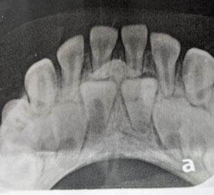
Emergency visit / exam + radiographs (bitewings or periapicals; if behavior limits imaging, clinician judgement).
-
Pain control: age-appropriate analgesics (follow local pediatric dosing or pharmacist/dentist guidance). Avoid benzocaine in very young children due to methemoglobinemia risk.
-
Infection control: antibiotics only if systemic signs (fever, spreading infection, cellulitis) or if the clinician judges it necessary. Antibiotics do not cure pulp disease — they are adjunctive.
-
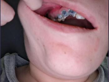
Arrest lesions fast: apply Silver Diamine Fluoride (SDF) to cavitated lesions to arrest decay and reduce pain — excellent temporizing measure in uncooperative/young children (discolors carious dentine black).
-
Temporary restorations / extraction decisions: restore teeth that are restorable (SSC after appropriate pulp therapy), extract non-restorable teeth.
-
If multiple teeth need extensive work or child cooperation is poor: plan treatment under general anesthesia (GA) with pediatric dental team.
-
Dietary counseling & oral hygiene: remove nocturnal bottle, reduce frequent sugary snacks, start brushing with age-appropriate fluoride toothpaste (pea-sized) with caregiver supervision.
-
Follow-up: definitive restorative/pulp therapy or extractions within 1–2 weeks after initial emergency measures.
Expected short outcomes in 14 days: acute pain/infection should be improved or controlled after emergency care, SDF can arrest many lesions quickly, soft tissue healing after extractions/pulpal surgery should be well underway. Carious tissue does not “heal” — arrested or removed.
Definitive treatment options (practical)
-
Restorable primary molars with pulp involvement → pulpotomy + stainless steel crown (SSC) or pulpectomy if necrotic.
-
Non-restorable primary molars → extraction with parental counseling and plan for space maintenance if needed.
-
Anterior primary teeth: if carious but restorable, consider composite or zirconia crowns for aesthetics; if non-restorable and extracted, assess speech/esthetic implications.
-
Multiple-tooth disease or young child → consider full-mouth rehabilitation under GA in pediatric dental center.
-
Prevention program: fluoride varnish at each visit, caregiver education, caries risk management, possible topical antimicrobials (chlorhexidine rinses not routine for very young children).
What will scale up if untreated
-
Increasing pain and sleepless nights; difficulty eating, weight loss or poor nutrition.
-
Acute dental abscesses, cellulitis, facial space infections — in severe cases can threaten airway or become systemic.
-
Spread to permanent tooth germs — enamel hypoplasia, delayed eruption or pathologic effects on successors.
-
Early tooth loss → space loss, future malocclusion, speech and mastication problems.
-
Behavioral/psychosocial effects from chronic pain and poor oral appearance.
Practical comments for caregivers (clear, short)
-
Do not wait — this is advanced disease. Call a pediatric dentist now.
-
For immediate relief: avoid very hot/cold foods, give age-appropriate pain reliever per label/prescriber, apply cold compress externally for swelling.
-
Start strict elimination of sugary drinks/snacks, remove bottle/juice at night, brush twice daily with fluoride paste under supervision.
Suggested workflow for the treating dentist (stepwise)
-
Triage (phone) → urgent appointment if systemic signs.
-
Exam + radiographs + assess behavior.
-
Apply SDF to arrest lesions now (consent for staining).
-
Plan definitive care: pulpotomy+SSC vs extraction vs GA full-mouth rehab.
-
Post-op preventive plan: fluoride varnish schedule, diet plan, recall every 3 months initially.
Timeframes (summary)
-
Emergent evaluation: within 48–72 hours.
-
Acute infection control / temporary arrest (SDF, analgesics, antibiotics if needed): within 0–7 days.
-
Definitive treatment (restorations, pulpotomy, extractions or GA rehab): ideally within 1–2 weeks of triage.
-
Soft tissue healing after extractions: 7–14 days.
-
Full recovery and restorative finish: depends on treatment plan; could be 1 appointment (GA) or several appointments over weeks.
Referral & location
Refer to a pediatric dentist or hospital dental clinic. For clinicians or caregivers in Cebu area, use the directory you provided:
https://cebudentalimplants.com/map-dental-clinic
Teeth Case
 |
 |
 |
 |
 |
 |