Where Bright Smiles Begin.

Hidden Tooth Infections: Silent Tooth Infection - A Japan-Focused Dental Case Study and Clinical Analysis
image hero:
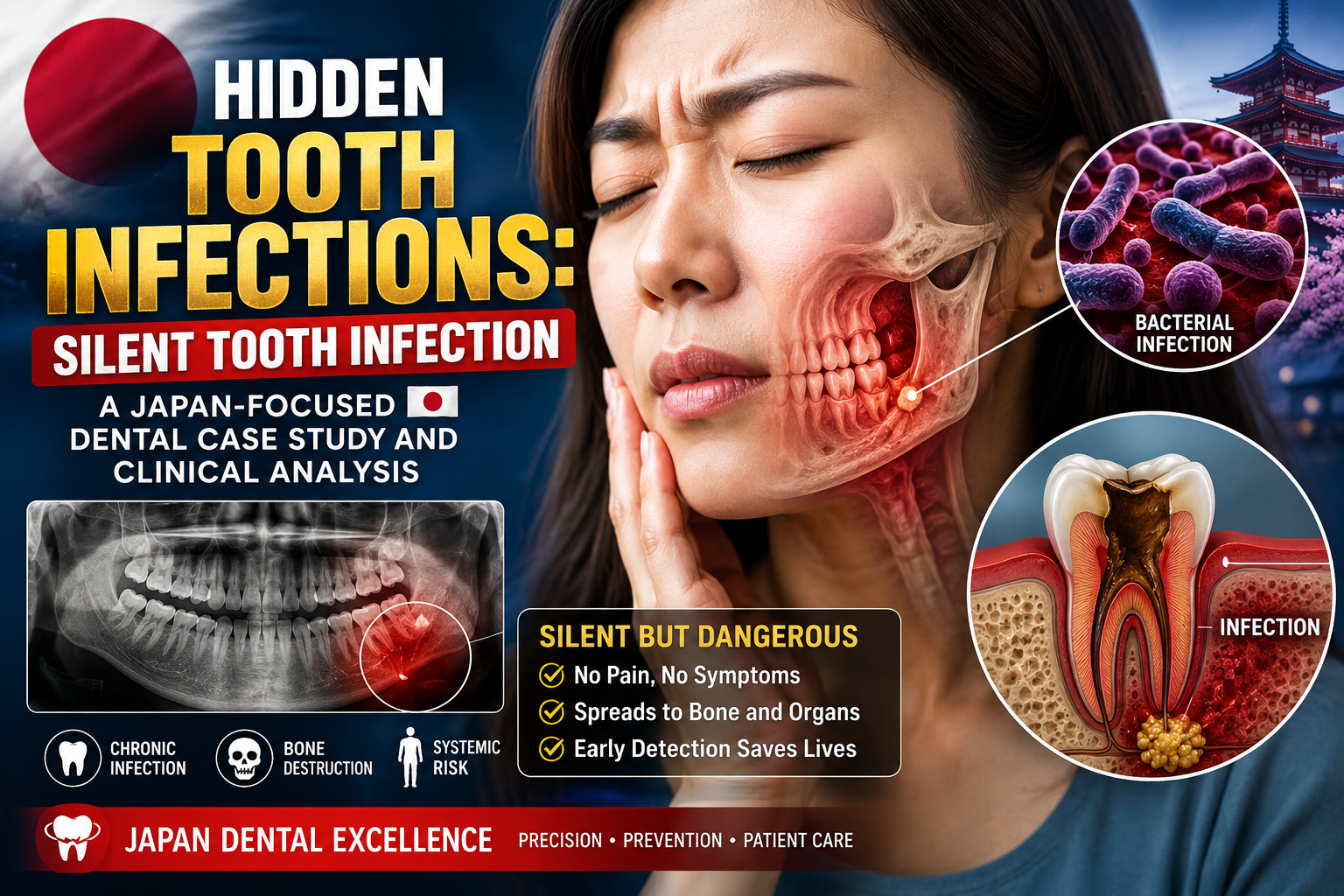
A tooth can look normal, feel normal, and still contain a serious underlying infection.
This is one of the most deceptive situations encountered in endodontic dentistry: the silent or hidden tooth infection. Patients may have little or no pain because the dental pulp has already lost vitality, while inflammation continues around the root and gradually affects the surrounding bone.
In clinical terminology, one important example is asymptomatic apical periodontitis—inflammation and destruction around the root apex that may produce a radiographic lesion without causing noticeable symptoms. A different presentation, chronic apical abscess, may also produce surprisingly little discomfort and can sometimes drain through a small sinus tract.
From a Japan-focused dental perspective, the key lesson is simple:
Absence of toothache does not prove absence of dental disease.
A comprehensive examination must combine the patient's history, clinical testing, periodontal evaluation, pulp sensibility testing, and appropriate dental imaging.
1. What Is a Hidden Tooth Infection?
A hidden tooth infection is not a single formal diagnostic category. It is a patient-friendly description for an infection or inflammatory process associated with a tooth that may have few or no obvious symptoms.
Common underlying conditions include:
-
Pulp necrosis
-
Asymptomatic apical periodontitis
-
Chronic apical abscess
-
Persistent infection after previous root canal treatment
-
Missed root canals
-
Coronal leakage or recurrent decay
-
Root fractures
-
Endodontic-periodontal lesions
When bacteria enter the pulp and the pulp subsequently becomes necrotic, microorganisms and inflammatory products can move through the root canal system toward the tissues surrounding the root.
Inflammation can then develop around the root apex and cause gradual destruction of supporting bone.
The patient may feel nothing because the nerve inside the tooth is no longer functioning normally.
2. Why Can a Tooth Infection Be Silent?
The common misconception
Many patients assume:
“If there is no pain, there is no infection.”
That assumption is incorrect.
Pain requires an appropriate biological pathway for the patient to perceive the inflammatory process. In a necrotic tooth, the pulp may no longer respond normally to thermal or electrical testing.
Meanwhile, microorganisms and inflammatory tissue can remain around the root.
The infection therefore may become a radiographic discovery rather than a pain-driven diagnosis.
The American Association of Endodontists describes asymptomatic apical periodontitis as an inflammatory and destructive process of pulpal origin that can appear as an apical radiolucency without clinical symptoms.
3. Case Study: The Patient Who Had No Toothache
Educational composite case
Consider a hypothetical patient in Japan in their early 50s.
The patient visits a dental clinic for a routine examination.
There is:
-
No spontaneous toothache
-
No facial swelling
-
No fever
-
No obvious pus
-
No major sensitivity to cold
-
Normal-looking gum tissue around most teeth
The patient says:
“I don't have any problems with this tooth.”
However, the dentist notices that a previously restored molar has an unusual clinical history.
The tooth had undergone extensive restorative treatment several years earlier.
A periapical radiograph is taken.
The image reveals a darkened area surrounding the root apex.
The patient is surprised.
“But it doesn't hurt.”
This is exactly where the diagnostic process becomes important.
The absence of pain does not eliminate the possibility of pulpal necrosis or apical disease.
4. Clinical Investigation
A careful endodontic examination should not depend on a single observation.
Step 1 — Patient history
The dentist asks:
-
Has the tooth ever been painful?
-
Was there previous trauma?
-
Was there a large filling?
-
Was a crown placed?
-
Was root canal treatment performed?
-
Has the tooth ever swollen?
-
Has a small pimple appeared on the gum?
-
Is there discomfort during chewing?
-
Has the patient experienced recurrent sinus problems on one side?
Previous symptoms may have disappeared after the pulp became necrotic.
Step 2 — Visual examination
The dentist examines:
-
Tooth discoloration
-
Existing restorations
-
Cracks
-
Recurrent decay
-
Crown margins
-
Gum swelling
-
Sinus tracts
-
Localized drainage
A tiny gum lesion can sometimes represent a drainage pathway from a chronic endodontic infection.
Chronic apical abscesses may have gradual onset with little or no discomfort and intermittent drainage through a sinus tract.
5. Pulp Sensibility Testing
One of the most important questions is:
Is the dental pulp still responding?
The dentist may use:
-
Cold testing
-
Heat testing when appropriate
-
Electric pulp testing
-
Comparison with neighboring and contralateral teeth
A nonresponsive tooth can raise suspicion of pulp necrosis, but a diagnosis should not be made from one test alone.
The result must be interpreted together with the patient's history, clinical examination, and imaging.
6. Percussion and Palpation
The dentist may gently test the tooth and surrounding tissues.
The tooth may be:
-
Non-tender
-
Mildly tender
-
Clearly painful to percussion
-
Tender to palpation
A silent infection can remain completely comfortable during these tests.
That is why a normal clinical response does not automatically exclude an underlying lesion.
7. Periodontal Probing
Periodontal probing provides another important diagnostic clue.
The dentist measures the depth around the tooth.
A generalized periodontal problem may produce multiple areas of increased probing depth.
A localized, unusually deep periodontal defect can raise different diagnostic questions, including:
-
Endodontic drainage
-
Root fracture
-
Combined endodontic-periodontal disease
-
Localized periodontal destruction
The distinction matters because these conditions can resemble one another clinically.
The AAE emphasizes that sensibility testing, percussion-related findings, transillumination, and periodontal probing can help differentiate endodontic and periodontal lesions.
8. The X-Ray: Where the Hidden Infection May Become Visible
The first major clue may be a periapical radiograph.
A dark area around the root apex can indicate loss or alteration of normal bone density associated with apical inflammation.
However, an important clinical principle is:
A radiograph alone does not establish the complete diagnosis.
Several different pathological processes can produce similar radiographic appearances, and definitive histologic differentiation may not be possible without tissue examination.
Therefore, the dentist correlates the image with clinical findings.
9. When CBCT Becomes Important
In selected cases, three-dimensional imaging such as limited-field CBCT can provide additional information.
It may help the clinician investigate:
-
Root anatomy
-
Missed canals
-
Periapical bone destruction
-
Cortical plate involvement
-
Root resorption
-
Treatment complications
-
Possible root fractures
-
Relationship to anatomical structures
CBCT should not automatically replace conventional radiographs.
It is generally used when the additional three-dimensional information can contribute meaningfully to diagnosis or treatment planning.
Recent AAE case examples demonstrate how limited-FOV CBCT can reveal apical lesions and anatomical findings that influence endodontic treatment decisions.
10. Case Analysis: What Was Actually Happening?
Returning to our educational case:
The patient had no significant pain.
The tooth appeared clinically quiet.
But the investigation showed:
Pulp testing:
Nonresponsive compared with appropriate control teeth.
Percussion:
Minimal or no discomfort.
Palpation:
Minimal or no discomfort.
Radiograph:
Periapical radiolucency associated with the tooth.
Clinical interpretation:
Findings consistent with pulpal necrosis and asymptomatic apical periodontitis, subject to confirmation by the treating dentist.
This is the classic pattern of a silent endodontic disease process.
11. Another Hidden Pattern: Chronic Apical Abscess
Not every silent infection looks exactly the same.
A second important pattern is a chronic apical abscess.
The patient may report:
“Sometimes I notice a small bump on my gum, but it doesn't hurt.”
The bump may disappear and return.
Occasionally, fluid or pus drains through the sinus tract.
Because the infection has an outlet, pressure may be reduced and the patient may experience surprisingly little pain.
That does not mean the underlying disease has disappeared.
A chronic apical abscess is characterized by gradual onset, little or no discomfort, and possible intermittent drainage through an associated sinus tract.
12. Why Waiting for Pain Can Be a Mistake
One of the most important educational messages for patients is:
Pain is not a reliable screening test for dental infection.
A tooth can become severely compromised before the patient experiences significant symptoms.
Possible warning clues include:
-
A tooth that no longer reacts normally to cold
-
A small recurring gum pimple
-
Unexplained drainage
-
Dark discoloration
-
Previous trauma
-
A large restoration
-
Previous root canal treatment
-
Pain that suddenly disappeared after a period of severe toothache
-
A radiographic lesion discovered during routine examination
A patient with any of these findings should receive a professional dental evaluation.
13. The Japan Clinical Perspective
In Japan, preventive and routine dental examinations can provide an important opportunity to detect disease before it becomes painful.
For a patient who says:
“Doctor, I don't have any pain.”
the appropriate response is not necessarily:
“Then everything is fine.”
Instead, the clinician should determine whether the tooth is actually healthy through appropriate examination.
This is particularly important for teeth with:
-
Large restorations
-
Crowns
-
Previous root canal treatment
-
Previous trauma
-
Deep caries
-
Suspicious radiographic findings
The clinical objective is not simply to find infection.
It is to determine what disease process is present, which tooth is responsible, whether the tooth can be preserved, and what treatment is appropriate.
14. Treatment: What Happens After Diagnosis?
Treatment depends entirely on the diagnosis.
For a restorable tooth with an infected or necrotic pulp, treatment may involve root canal treatment.
The objective is to:
-
Gain access to the canal system.
-
Locate the canals.
-
Remove infected and necrotic contents.
-
Clean and shape the canal system.
-
Disinfect the system.
-
Seal the canals appropriately.
-
Restore the tooth to protect it from further damage.
-
Monitor healing.
The AAE notes that endodontic treatment for an abscessed tooth involves cleaning and shaping the root canals and sealing the space, followed by definitive restoration.
15. What If the Tooth Was Already Root-Canal Treated?
This is a more complicated situation.
A previously treated tooth can develop persistent or recurrent apical disease.
Potential causes include:
-
Missed anatomy
-
Persistent microbial infection
-
Inadequate previous treatment
-
Coronal leakage
-
Root fracture
-
Perforation
-
Complex canal anatomy
-
New infection
The correct response is not automatically extraction.
The clinician must determine why the previous treatment failed.
AAE case reports demonstrate that complex previously treated teeth with asymptomatic apical disease or chronic abscess can require detailed diagnostic assessment and may sometimes be managed with retreatment or surgical approaches.
16. Antibiotics Are Not Automatically the Solution
This is an important distinction.
A hidden chronic infection should not simply be treated by taking antibiotics without addressing the dental source.
When appropriate, definitive dental treatment may involve:
-
Root canal treatment
-
Endodontic retreatment
-
Endodontic surgery
-
Drainage
-
Extraction when the tooth cannot predictably be saved
Antibiotics have specific indications and are not a substitute for eliminating the source of an endodontic infection.
The AAE notes that many localized chronic apical abscesses do not require systemic antibiotic therapy when appropriate local dental treatment can be provided, while systemic involvement or spreading infection changes the urgency and management.
17. When a “Silent” Infection Becomes an Emergency
The word silent should never be interpreted as harmless.
Seek urgent professional assessment if a dental infection is associated with:
-
Rapidly increasing facial swelling
-
Significant jaw swelling
-
Fever
-
Malaise
-
Difficulty swallowing
-
Difficulty breathing
-
Increasing difficulty opening the mouth
-
Spreading facial or neck swelling
-
Significant systemic illness
Rapidly spreading infection and systemic involvement require a different level of urgency.
If breathing or swallowing is compromised, emergency medical evaluation is appropriate.
18. A Special Diagnostic Challenge: Upper Teeth and the Maxillary Sinus
A hidden dental infection can occasionally be associated with sinus problems.
The roots of some upper posterior teeth are anatomically close to the maxillary sinus.
Apical inflammation can affect the adjacent sinus mucosa, and dental disease can sometimes be overlooked when a patient presents primarily with unilateral or recurrent sinus symptoms.
The AAE notes that endodontic-origin maxillary sinus disease can be difficult to recognize radiographically and requires careful clinical assessment of the involved teeth.
This creates an important diagnostic question:
Is the problem actually coming from the sinus—or from a tooth next to the sinus?
19. Expert Case Discussion
Case finding
A patient presents without tooth pain.
Initial assumption
“No symptoms, therefore low concern.”
Diagnostic examination
A previously restored molar shows:
-
Abnormal pulp sensibility
-
No significant percussion pain
-
No major swelling
-
Periapical radiolucency
Diagnostic shift
The case moves from:
“Healthy tooth with no symptoms”
to:
“Potential pulpal necrosis with asymptomatic apical disease.”
Treatment planning
The dentist must then determine:
-
Is the tooth restorable?
-
Is the infection endodontic?
-
Is there a periodontal component?
-
Is there a crack or root fracture?
-
Was the tooth previously treated?
-
Is retreatment possible?
-
Is surgery indicated?
-
Would extraction provide a better prognosis?
This is where clinical dentistry becomes more than simply looking for pain.
20. The Most Important Lesson
A hidden tooth infection is a diagnostic problem before it becomes a pain problem.
The patient may feel healthy.
The tooth may look normal.
The gum may appear normal.
But the root and surrounding bone may tell another story.
The correct approach is not to frighten patients into unnecessary treatment.
The correct approach is objective diagnosis.
A dentist should correlate:
History + Clinical Examination + Pulp Testing + Periodontal Probing + Radiographs + CBCT when indicated
before making a treatment decision.
21. Patient Takeaway
If you have no toothache, that is good—but it does not guarantee that every tooth is healthy.
Consider a dental examination if you have:
-
A tooth with a large old filling
-
A previous root canal
-
A history of dental trauma
-
A recurring gum bump
-
Unexplained drainage
-
A tooth that behaves differently from neighboring teeth
-
A suspicious finding on an X-ray
-
Recurrent one-sided sinus symptoms
-
A tooth that suddenly stopped hurting after previously severe pain
Early diagnosis may allow the dentist to evaluate whether the tooth can be preserved before the problem becomes more destructive.
Conclusion
Hidden Tooth Infections, Silent Tooth Infections, and asymptomatic apical disease demonstrate why dentistry cannot rely on pain alone.
A chronic infection may progress quietly around the root of a tooth while the patient continues eating, speaking, and living normally.
The most important clinical question is therefore not:
“Does it hurt?”
It is:
“What does the complete examination show?”
For patients in Japan and elsewhere, routine dental evaluation, appropriate radiographic assessment, and careful endodontic diagnosis can reveal disease that would otherwise remain invisible.
A silent tooth infection is not a diagnosis to make from symptoms alone. It is a condition that requires professional examination to identify its source, severity, and appropriate treatment.
Educational disclaimer: This case study is a fictional composite created for dental education and does not represent an individual Japanese patient. Treatment decisions must be made by a licensed dentist or endodontic specialist after an appropriate clinical examination.
Related Clinical Cases
Patients with lower front gum inflammation may also experience tartar buildup, periodontal disease, gum abscesses, or tooth decay. Explore these related clinical case studies.



Other topics:
Other Dental Clinic
 |
 |
 |
 |





