Because Every Tooth Deserves Care.

Internal Root Resorption: A USA Dentist’s Zoom-In Clinical Review, Case Analysis, Diagnosis, and Treatment Advice
Internal root resorption is one of the more unusual problems an endodontist encounters because the tooth can begin destroying its own internal dentin structure from the inside.
Unlike ordinary tooth decay, which generally starts from the external tooth surface, internal root resorption begins within the pulp/root-canal space and can progressively enlarge the internal canal by destroying surrounding dentin. The American Association of Endodontists describes internal resorption as a pathologic loss of dentin associated with inflammatory activity within the pulp/root-canal system.
The clinical challenge is that the condition may remain silent for a long period.
By the time the patient notices discoloration, pain, a small swelling, or a dentist discovers an unusual radiographic finding, significant structural damage may already have occurred.
1. Zoom-In: What Is Actually Happening Inside the Tooth?
Imagine taking a microscopic camera and moving through the tooth from the crown toward the root.
Zoom Level 1 — Healthy Tooth
A healthy tooth has:
-
Enamel
-
Dentin
-
Pulp tissue
-
Root canal
-
Cementum
-
Periodontal ligament
-
Supporting bone
The dentin forms the strong internal body of the tooth around the pulp.
Zoom Level 2 — Pulp Inflammation
Internal root resorption is associated with inflammation and damage to the protective predentin lining of the pulp/root canal.
This inflammatory environment can activate specialized cells capable of resorbing mineralized dental tissues.
Zoom Level 3 — Dentin Begins Disappearing
The resorptive process progressively removes dentin from the inside.
Instead of the tooth being attacked from the outside, the internal canal space becomes enlarged.
Zoom Level 4 — Structural Weakening
As more dentin disappears, the remaining root wall can become dangerously thin.
At this stage, the tooth may become vulnerable to:
-
Root perforation
-
Fracture
-
Infection
-
Loss of structural integrity
Internal root resorption has been described radiographically as a characteristic enlargement or "ballooning" of the canal space.
2. Why Patients Often Do Not Know They Have It
One of the most important clinical lessons is:
Internal root resorption can be asymptomatic.
A patient may feel completely normal while the lesion progresses.
A review of root resorption literature notes that root resorption is often discovered incidentally during radiographic examination. Possible later manifestations include pain, discoloration, and tooth mobility.
This is why a routine dental examination can occasionally uncover a major problem that the patient never suspected.
3. USA Case Study: The Silent Incisor
Illustrative Clinical Case
Consider a hypothetical 34-year-old patient who visits a U.S. dental office for a routine examination.
The patient reports:
"I don't have any pain. I just want my teeth checked."
During the examination, the dentist notices that one upper front tooth appears slightly darker than the neighboring teeth.
There is no major swelling.
There is no obvious cavity.
The tooth does not appear dramatically damaged from the outside.
A periapical radiograph is obtained.
Initial Zoom-In Finding
The radiograph reveals an unusual, rounded enlargement within the root-canal space.
The dentist becomes concerned about internal root resorption.
The patient is then referred to an endodontist for further evaluation.
4. The Endodontic Zoom-In
The endodontist evaluates the tooth from several perspectives.
Clinical Examination
The dentist assesses:
-
Tooth color
-
Tenderness
-
Mobility
-
Periodontal probing
-
Pulp status
-
Periapical tissues
-
Adjacent teeth
Periapical Radiograph
The radiograph can reveal the characteristic internal enlargement of the canal.
But a two-dimensional image does not always tell the complete three-dimensional story.
CBCT Evaluation
When clinically justified, cone-beam computed tomography can provide three-dimensional information about:
-
Lesion location
-
Lesion size
-
Remaining dentin
-
Root anatomy
-
Possible perforation
-
Relationship to surrounding structures
Research and clinical reviews have emphasized the contribution of CBCT to diagnosing and planning treatment for root-resorption cases.
Importantly, CBCT should not simply be ordered automatically for every dental patient. The imaging decision should be based on the clinical question and expected diagnostic benefit.
5. Internal vs. External Root Resorption
This distinction is critical.
Internal Root Resorption
The process originates from within the pulp/root-canal system.
The lesion typically expands the internal dentin.
External Root Resorption
The resorptive process begins from the external root surface or periodontal tissues.
These conditions can sometimes look similar on conventional radiographs.
The American Association of Endodontists emphasizes that root resorption is not simply one disease divided into "internal" and "external"; there are multiple resorptive entities that require careful differential diagnosis.
This is why an unusual radiographic lesion should not automatically be labeled internal resorption without appropriate evaluation.
6. What Causes Internal Root Resorption?
The exact biology is complex, and not every case has an identifiable trigger.
Potential associations reported in the literature include:
-
Dental trauma
-
Pulpal inflammation or infection
-
Previous dental procedures
-
Orthodontic treatment
-
Other forms of injury to the tooth
A recent review found trauma among the commonly reported associations, along with orthodontic movement, pulpotomy procedures, pulp infection, and other factors.
However, patients should understand an important point:
Sometimes the precise cause cannot be determined.
It is therefore inappropriate to automatically blame the patient for the condition.
The AAE also notes that resorption can sometimes occur without an obvious reason.
7. The "Pink Tooth" Warning Sign
One fascinating clinical presentation is the "Pink Tooth of Mummery."
A tooth affected by internal resorption may sometimes develop a pink or reddish appearance because the resorptive process can bring vascularized tissue closer to the crown.
But a pink tooth is not diagnostic by itself.
Pink discoloration can have other causes, and modern literature emphasizes that this clinical sign is not exclusive to internal root resorption.
Therefore:
Pink tooth = investigate.
It does not automatically mean:
Pink tooth = internal resorption.
8. Why Internal Resorption Is Dangerous
The greatest concern is progressive loss of dentin.
Think of the root as a structural tube.
The dentin provides much of the mechanical strength of that tube.
If internal resorption removes substantial amounts of dentin, the remaining wall can become very thin.
Eventually, the lesion may communicate with the external root surface.
This is called perforating internal resorption.
A perforation can significantly complicate treatment because the dentist now has to control the defect and establish a durable seal.
9. Treatment: Can the Tooth Be Saved?
In many cases, yes.
The primary objective is to stop the biological process and eliminate the inflamed/infected tissue that sustains it.
Conventional root canal treatment remains the principal treatment approach for internal root resorption in appropriately treatable teeth. The procedure removes the pulp tissue and blood supply that support the resorptive process.
The earlier the lesion is detected and treated, the more favorable the structural situation may be.
10. Root Canal Treatment — Zoom-In
During treatment, the endodontist carefully accesses the root canal.
The goal is to:
-
Locate the canal system.
-
Remove diseased tissue.
-
Disinfect the canal.
-
Clean the resorptive cavity as effectively as possible.
-
Seal the canal and resorptive defect.
-
Restore the tooth to protect its remaining structure.
Advanced cases may require specialized techniques, magnification, ultrasonic irrigation, and biocompatible repair materials.
Calcium-silicate materials such as MTA may be used in selected perforating defects to establish a biocompatible seal.
11. When the Root Has Been Perforated
This is where treatment becomes considerably more complex.
The dentist must determine:
Where is the perforation?
How large is it?
Is the tooth restorable?
Is there adequate remaining tooth structure?
Is the periodontal attachment compromised?
Can the defect be sealed predictably?
A perforation does not automatically mean the tooth must be extracted.
Published clinical reviews report that conservative treatment can sometimes achieve good outcomes even in selected perforating internal-resorption cases.
However, prognosis is highly case-dependent.
12. 2026 Case Evidence: What New Data Tell Us
A 2026 retrospective study from Tufts University School of Dental Medicine examined 50 internal-resorption cases evaluated with periapical radiographs and CBCT.
The study reported that 98% of the cases were classified as internal inflammatory resorption and 2% as internal replacement resorption. Perforation was present in 36% of the cases.
This is clinically important because it demonstrates how variable these lesions can be.
It also reinforces why a simple two-dimensional image may not provide enough information for complicated cases.
13. When Extraction May Become Necessary
Not every resorbed tooth can be saved.
Extraction may be considered when:
-
Remaining tooth structure is inadequate
-
The root is severely compromised
-
The tooth cannot be predictably restored
-
The lesion has caused extensive structural destruction
-
There is an unfavorable fracture
-
Infection or periodontal destruction cannot be controlled
-
The long-term prognosis is extremely poor
The objective should never be:
"Save every tooth at any cost."
The objective is:
"Determine whether this tooth can be predictably maintained."
14. What Happens After Extraction?
If the tooth cannot be saved, the patient may consider replacement options such as:
-
Dental implant
-
Fixed dental bridge
-
Removable partial denture
-
Other prosthetic options
If an implant is being considered, the clinician must evaluate the remaining bone and soft tissue after extraction.
The treatment plan should therefore consider the entire sequence:
Diagnosis → Endodontic treatment or extraction → Healing → Structural restoration → Long-term maintenance
15. USA Dentist's Clinical Advice
If your dentist tells you that you have internal root resorption, don't immediately assume that the tooth is hopeless.
Instead, ask:
Question 1
Where exactly is the resorption located?
Question 2
How much dentin remains?
Question 3
Has the root been perforated?
Question 4
Is the pulp still vital or infected?
Question 5
Do I need CBCT imaging for treatment planning?
Question 6
Can an endodontist evaluate this tooth?
Question 7
Can the tooth be restored after root canal treatment?
Question 8
What is the expected prognosis?
Question 9
What happens if we attempt to save the tooth?
Question 10
What is the alternative if the tooth cannot be predictably restored?
16. Dental Hub Review: The Most Important Lesson
Internal root resorption is a rare and potentially destructive dental condition that can remain hidden.
The patient may have:
Little pain + minimal visible damage + significant internal destruction.
That combination is precisely why radiographic evaluation and appropriate endodontic assessment can be important.
The American Association of Endodontists describes early detection, removal of the cause, and appropriate endodontic treatment as central to management.
From a USA endodontic perspective, I would summarize the clinical strategy this way:
Find it early. Map it accurately. Determine whether the tooth is restorable. Remove the diseased tissue. Seal the defect. Protect the remaining tooth structure.
The goal is not simply to perform a root canal.
The goal is to stop the resorptive process while preserving as much healthy tooth structure as possible and maintaining a predictable long-term restoration.
Important Patient Disclaimer
This case analysis is educational and uses an illustrative clinical scenario rather than identifying a real patient. Internal root resorption requires individualized diagnosis by a dentist or endodontist. Radiographs and, when clinically justified, CBCT may be used to determine the location and extent of the lesion. Treatment decisions depend on the tooth's remaining structure, periodontal condition, restorability, and overall clinical findings.
Related Clinical Cases
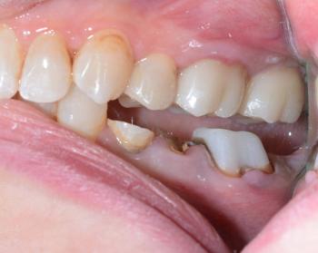
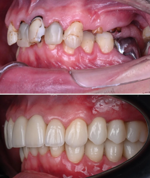
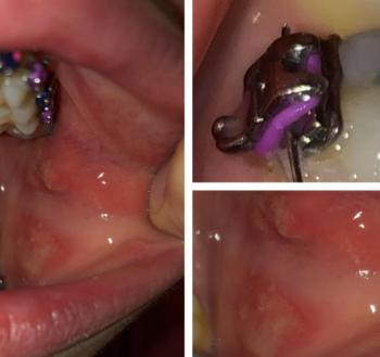
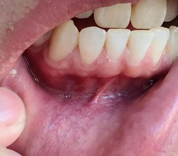
Patients with lower front gum inflammation may also experience tartar buildup, periodontal disease, gum abscesses, or tooth decay. Explore these related clinical case studies.
Other topics:
Other Dental Clinic