Because Every Tooth Deserves Care.

Lower Front Gum Inflammation Case Analysis and Healing Timeline
Image:
Severity:
Teeth Problems:
Teeth Case – Full Analysis and Provisional Diagnosis (Zoom 100%)
This assessment is based on a single photographic image. A definitive diagnosis requires an in-person dental examination, periodontal probing, and radiographs if indicated.
1. Visual Findings (Deep Examination)
-
Teeth condition:
-
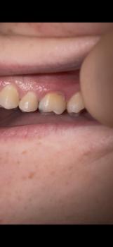
No obvious cavities or fractures visible.
-
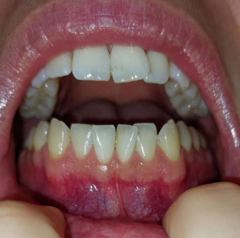
Mild yellowish deposits along the cervical (neck) areas of the lower front teeth, consistent with plaque or early calculus (tartar) buildup.
-
-
Gingival condition (gums):
-
Pronounced redness and inflammation along the lower anterior gingival margin.
-
Mild swelling of gum tissue, suggesting active inflammation.
-
-
Oral hygiene indicators:
-
Pattern indicates plaque retention at the gumline rather than deep structural tooth damage.
-
-
Occlusion:
-
Bite appears acceptable with no obvious traumatic contact in this image.
-
2. Most Likely Diagnosis (Provisional)
-
Plaque-induced gingivitis (lower anterior region predominant)
-
Early periodontal inflammation (reversible stage)
-
Localized calculus accumulation
3. Issues That May Scale Up if Untreated
If left untreated for 14–30 days, the following may occur:
-
Progression from gingivitis to early periodontitis
-
Persistent gum bleeding
-
Gum recession exposing tooth roots
-
Increased tooth sensitivity
-
Formation of periodontal pockets
-
Long-term risk of bone loss and tooth mobility
4. Recommended Treatment Process
Phase 1: Professional Care (Days 1–3)
-
Comprehensive dental examination
-
Periodontal probing of lower anterior teeth
-
Professional scaling and polishing to remove plaque and calculus
-
Oral hygiene instruction
Phase 2: Home Inflammation Control (Days 4–7)
-
Twice-daily brushing with soft-bristled toothbrush
-
Focused cleaning along the gumline
-
Daily flossing or interdental brush use
-
Antimicrobial mouth rinse if recommended by dentist
Phase 3: Re-evaluation (Days 8–14)
-
Visible reduction in redness and swelling expected
-
Bleeding should significantly decrease
-
Further periodontal treatment planned only if inflammation persists
Expected healing time:
-
Gingival inflammation typically improves within 7–14 days with proper plaque control.
5. If Healing Takes 14 Days – What May Scale Up
-
Persistent gingival inflammation
-
Early attachment loss
-
Need for deeper periodontal cleaning
-
Increased maintenance frequency
-
Higher long-term periodontal risk
6. Home Care Recommendations (Interim)
-
Brush gently but thoroughly along the gumline
-
Avoid aggressive brushing that may worsen gum recession
-
Floss daily
-
Avoid smoking
-
Reduce sugary and acidic food intake
7. Professional Commentary
This case shows healthy tooth structure with unhealthy gums, a common and fully reversible condition when treated early. Prompt plaque removal and consistent oral hygiene can restore gum health and prevent progression to periodontal disease.
What Is Happening to the Gums?
The teeth in this case look healthy and strong, but the gums around the lower front teeth are red and swollen. This condition is called gingivitis. Gingivitis is an early and common form of gum disease caused by plaque buildup along the gumline.
The good news is that gingivitis is reversible when treated early.
What Causes Red and Inflamed Gums?
Gum inflammation usually develops when plaque is not fully removed during daily brushing and flossing. Common causes include:
-
Incomplete brushing along the gumline
-
Not flossing regularly
-
Plaque hardening into tartar
-
Smoking or vaping
-
Poor oral hygiene habits
Even if teeth look clean, plaque can still collect where the teeth meet the gums.
Is This a Serious Problem?
At this stage, the condition is not serious and not permanent. However, if left untreated, gingivitis can progress into periodontitis, a more severe gum disease that can cause:
-
Gum recession
-
Bone loss
-
Tooth sensitivity
-
Loose teeth
Early treatment prevents these problems.
Recommended Treatment Process
Step 1: Professional Dental Cleaning (Days 1–3)
A dentist or dental hygienist will:
-
Examine the gums
-
Remove plaque and tartar through professional scaling
-
Polish the teeth to slow plaque buildup
This step is essential because hardened plaque cannot be removed at home.
Step 2: Home Care and Healing (Days 4–7)
After cleaning, patients should:
-
Brush twice daily using a soft toothbrush
-
Clean gently along the gumline
-
Floss once daily
-
Use a dentist-recommended mouth rinse if advised
Redness and swelling usually begin to improve during this period.
Step 3: Re-evaluation (Days 8–14)
Within 1–2 weeks:
-
Gum color should return closer to pink
-
Swelling should reduce
-
Bleeding should stop or greatly decrease
If inflammation remains, further gum treatment may be needed.
How Long Does Healing Take?
With proper treatment and home care:
-
7–14 days for noticeable gum healing
-
Full improvement depends on daily oral hygiene habits
What Happens If Treatment Is Delayed?
If gingivitis is ignored for several weeks or months, it may lead to:
-
Persistent gum bleeding
-
Gum recession
-
Periodontal pockets
-
Bone loss around teeth
-
Higher treatment costs later
Early care is always simpler and more affordable.
What You Can Do at Home
To keep gums healthy:
-
Brush gently along the gumline
-
Floss daily
-
Avoid aggressive brushing
-
Avoid smoking
-
Visit your dentist regularly
Consistency is more important than brushing hard.
Frequently Asked Questions (FAQ): Localized Plaque-Induced Anterior Gingivitis
Reviewed by a USA Dental Expert
1. What is localized plaque-induced anterior gingivitis?
Localized plaque-induced anterior gingivitis is an early-stage gum disease caused by bacterial plaque accumulating around the gumline, most commonly affecting the lower front teeth. The condition causes redness, swelling, tenderness, and bleeding during brushing or flossing. At this stage, the infection is limited to the gums and has not damaged the bone or supporting tissues, making it completely reversible with appropriate treatment.
2. What are the most common symptoms?
Patients often notice:
- Red, swollen gums around the lower front teeth
- Bleeding while brushing or flossing
- Tenderness along the gumline
- Persistent bad breath (halitosis)
- Mild discomfort rather than severe pain
- Increased plaque or tartar near the gumline
Many patients experience little or no pain, which is why early gingivitis is frequently overlooked.
3. Why does gingivitis usually affect the lower front teeth first?
The lower front teeth are located immediately next to the openings of the submandibular and sublingual salivary glands.
These glands constantly release saliva that contains calcium and phosphate minerals. If plaque is not removed daily, these minerals rapidly harden the plaque into calculus (tartar), making this area one of the most common locations for tartar buildup in the mouth.
4. Can gingivitis heal on its own?
No.
Although the body's immune system attempts to control the inflammation, plaque and tartar must be physically removed before complete healing can occur.
Professional dental cleaning combined with excellent home care is necessary to eliminate the source of the inflammation.
5. Is plaque-induced gingivitis reversible?
Yes.
One of the most encouraging aspects of plaque-induced gingivitis is that it is completely reversible when treated early.
Once bacterial plaque and calculus are removed, healthy gum tissue usually returns without permanent damage.
6. How long does recovery usually take?
Most patients notice improvement within:
- 48–72 hours: swelling begins decreasing
- 5–7 days: bleeding significantly improves
- 7–14 days: gums often return to a healthy pink appearance
Healing depends on maintaining excellent oral hygiene every day.
7. What causes the gums to bleed during brushing?
Bleeding is a sign of inflammation.
Bacteria within dental plaque release toxins that trigger the immune system. Blood vessels inside the gums enlarge and become fragile, causing bleeding even with gentle brushing or flossing.
Healthy gums should not bleed during normal oral hygiene.
8. Is gingivitis painful?
Usually not.
Most patients experience only:
- Mild tenderness
- Gum sensitivity
- Slight swelling
Because severe pain is uncommon, many individuals delay seeking treatment until the condition becomes more advanced.
9. What happens during a professional dental cleaning?
A professional prophylaxis typically includes:
- Comprehensive oral examination
- Periodontal assessment
- Removal of plaque
- Removal of tartar (calculus)
- Tooth polishing
- Personalized brushing and flossing instruction
The procedure usually takes between 30 and 60 minutes, depending on the amount of plaque and calculus present.
10. Will I need antibiotics?
In most cases, no.
Simple plaque-induced gingivitis is treated by removing plaque and improving oral hygiene.
Antibiotics are generally reserved for more serious infections involving facial swelling, abscesses, or advanced periodontal disease.
11. Will I need a deep cleaning?
Not necessarily.
Deep cleaning (Scaling and Root Planing) is recommended only when periodontitis has developed with:
- periodontal pockets
- attachment loss
- bone loss
If the gums remain healthy underneath and pocket depths are normal, a routine professional cleaning is usually sufficient.
12. What is the difference between gingivitis and periodontitis?
| Gingivitis | Periodontitis |
|---|---|
| Gum inflammation only | Inflammation plus bone loss |
| Reversible | Irreversible tissue destruction |
| No attachment loss | Loss of supporting structures |
| No deep pockets | Periodontal pockets develop |
| Usually treated with prophylaxis | Often requires deep cleaning and ongoing periodontal care |
13. Can untreated gingivitis become serious?
Yes.
Without treatment, gingivitis may progress into periodontitis, which can lead to:
- gum recession
- deep periodontal pockets
- bone loss
- loose teeth
- eventual tooth loss
Early treatment helps prevent these complications.
14. How can I prevent plaque-induced gingivitis from returning?
Daily prevention includes:
- Brush twice daily using a soft-bristled toothbrush.
- Angle the bristles toward the gumline using the Modified Bass Technique.
- Floss every day.
- Clean between teeth with interdental brushes when appropriate.
- Limit sugary snacks and beverages.
- Visit your dentist every six months for professional cleanings.
- Replace your toothbrush every three to four months.
15. Is mouthwash enough to cure gingivitis?
No.
Mouthwash can reduce bacteria temporarily, but it cannot remove hardened tartar.
Mechanical plaque removal through brushing, flossing, and professional cleaning remains the foundation of successful treatment.
16. Why does tartar form so quickly?
Dental plaque can begin hardening into calculus within 24 to 72 hours, especially around the lower front teeth where saliva contains high concentrations of calcium.
Once tartar forms, it cannot be removed with brushing alone.
17. Does smoking increase the risk of gingivitis?
Yes.
Smoking:
- reduces blood flow to the gums
- weakens the immune response
- slows healing
- increases plaque accumulation
- significantly raises the risk of progression to periodontitis
Smokers may also have less noticeable bleeding despite having more advanced disease.
18. Can diabetes make gingivitis worse?
Yes.
Poorly controlled diabetes increases inflammation, slows healing, and makes gum infections more difficult to control.
Likewise, untreated periodontal inflammation can make blood sugar management more challenging.
19. How often should I have professional dental cleanings?
For most healthy adults, professional cleanings every six months are appropriate.
Patients with frequent tartar buildup, diabetes, smoking history, orthodontic appliances, or previous gum disease may benefit from more frequent maintenance visits as recommended by their dentist.
20. When should I schedule a dental appointment immediately?
Seek prompt dental evaluation if you experience:
- Bleeding gums lasting longer than one week despite good oral hygiene
- Persistent swelling
- Gum pain that worsens
- Pus around the gums
- Loose teeth
- Gum recession
- Persistent bad breath
- Fever or facial swelling
These symptoms may indicate progression beyond simple gingivitis and require comprehensive periodontal evaluation.
Expert Summary
Localized plaque-induced anterior gingivitis is one of the most common and treatable oral health conditions seen in dental practices across the United States. When diagnosed early, professional plaque removal combined with consistent home care typically restores healthy gums within one to two weeks. The key is not to ignore early warning signs such as bleeding during brushing or localized gum swelling. Prompt intervention helps prevent progression to periodontitis, preserving the health of your gums, supporting bone, and natural teeth for years to come.
Professional Advice
This case shows healthy teeth with unhealthy gums, which is very common. Gingivitis does not cause permanent damage if treated early. Regular dental cleanings and good home care can fully restore gum health.
Localized Plaque-Induced Gingivitis of the Lower Front Teeth: A USA Dental Expert Clinical Case Study and Evidence-Based Review
Bleeding gums during brushing are often dismissed as a minor inconvenience. However, from a periodontal perspective, gingival bleeding is one of the earliest clinical warning signs of inflammation caused by bacterial plaque. When diagnosed early, this condition is completely reversible. When ignored, it may progress to irreversible periodontal disease that threatens the supporting structures of the teeth.
This clinical case study from Cebu Dental Implants presents a classic example of localized plaque-induced gingivitis affecting the mandibular anterior region, supported by current periodontal principles used throughout the United States and consistent with recommendations from the American Academy of Periodontology (AAP), the American Dental Association (ADA), and peer-reviewed periodontal research.
Executive Clinical Summary
Patient Presentation
An adult patient presented with the following concerns:
- Bleeding while brushing the lower front teeth
- Mild gum swelling
- Localized redness near the gumline
- Occasional tenderness
- Persistent plaque accumulation despite routine brushing
The patient denied severe pain, facial swelling, loose teeth, trauma, or recent dental procedures.
Clinical Examination
Extraoral Findings
- No facial swelling
- No cervical lymphadenopathy
- Normal mandibular movement
- No signs of acute odontogenic infection
Intraoral Findings
Affected Region:
Lower anterior gingiva surrounding teeth #23–#26 (Universal Numbering System / FDI #31–41)
Clinical observations included:
- Pronounced marginal erythema
- Mild localized edema
- Bleeding upon gentle periodontal probing
- Visible supragingival plaque
- Early supragingival calculus deposits
- Loss of normal gingival stippling
- No gingival recession
- No periodontal pocketing greater than 3 mm
All affected teeth demonstrated:
- Intact enamel
- No cavitated caries
- No fractures
- No mobility
- Stable occlusion
Radiographic evaluation revealed no evidence of alveolar bone loss.
Provisional Diagnosis
Plaque-Induced Gingivitis on an Intact Periodontium
This diagnosis represents the earliest and most treatable stage of periodontal disease.
Because inflammation remains confined to the gingival tissues, the supporting periodontal ligament and alveolar bone remain completely intact.
With appropriate treatment, complete recovery is expected.
Why the Lower Front Teeth Are Especially Vulnerable
One of the most common questions patients ask is:
"Why does tartar always build up behind my lower front teeth?"
The answer lies in normal oral anatomy.
Directly beneath the tongue are the openings of the:
- Wharton's ducts (submandibular glands)
- Sublingual salivary ducts
These glands continuously release saliva rich in calcium and phosphate minerals.
When bacterial plaque remains on tooth surfaces, these minerals rapidly harden the plaque into dental calculus.
This mineralization process commonly begins within 24 to 72 hours, making the lingual surfaces of the lower incisors the most frequent site of tartar accumulation in the human mouth.
Minor crowding, narrow embrasures, and inadequate flossing further increase plaque retention in this region.
Biological Mechanism of Disease
Plaque-induced gingivitis develops through a well-understood inflammatory pathway.
As plaque accumulates, bacteria release toxins that stimulate the body's immune response.
This causes:
- Dilation of gingival blood vessels
- Increased blood flow
- Migration of inflammatory cells
- Swelling of connective tissue
- Increased gingival crevicular fluid
- Bleeding during brushing or periodontal probing
At this stage:
- No connective tissue attachment has been lost.
- No periodontal ligament destruction has occurred.
- No alveolar bone resorption is present.
Because structural support remains intact, the disease is fully reversible.
Differential Diagnosis
Although this patient demonstrated classic plaque-induced gingivitis, American clinicians also consider other causes of localized gingival enlargement when inflammation fails to resolve after appropriate therapy.
Drug-Induced Gingival Enlargement
Common medications include:
- Nifedipine
- Amlodipine
- Phenytoin
- Cyclosporine
These medications may produce firm, lobulated gingival overgrowth that persists despite excellent plaque control.
Pyogenic Granuloma
Persistent irritation from plaque or calculus may occasionally produce a localized hyperplastic lesion, especially during pregnancy.
Unlike simple gingivitis, pyogenic granulomas often require surgical removal after inflammation has been controlled.
Early Periodontitis
If clinical attachment loss or radiographic bone loss is detected, the diagnosis changes from gingivitis to periodontitis, requiring more extensive periodontal therapy.
Treatment Plan
Phase I – Professional Periodontal Therapy (Day 1)
The patient underwent:
- Comprehensive periodontal examination
- Full periodontal charting
- Ultrasonic supragingival scaling
- Hand instrumentation for residual calculus removal
- Professional polishing
- Oral hygiene instruction
The patient received individualized instruction on the Modified Bass brushing technique, emphasizing plaque removal along the gingival margin.
Phase II – Home Care (Days 1–7)
The patient was instructed to:
- Brush twice daily using a soft-bristled toothbrush.
- Hold the brush at a 45-degree angle toward the gumline.
- Use gentle vibratory motions rather than aggressive scrubbing.
- Floss daily to disrupt interdental plaque.
- Use an alcohol-free chlorhexidine 0.12% mouth rinse if prescribed.
Smoking cessation and reduction of sugary beverages were also recommended.
Phase III – Clinical Re-evaluation (Days 7–14)
At follow-up, clinicians expect:
- Significant reduction in redness
- Resolution of swelling
- Elimination of bleeding on probing
- Return of healthy stippled gingival texture
- Improved plaque control
Most uncomplicated plaque-induced gingivitis resolves completely within 7 to 14 days when professional cleaning is combined with effective home care.
Comparison with Published USA Clinical Evidence
| Clinical Parameter | Cebu Clinical Case | Standard USA Clinical Findings |
|---|---|---|
| Primary Cause | Plaque and supragingival calculus | Microbial biofilm with calculus accumulation |
| Common Site | Lower front teeth | Mandibular anterior teeth |
| Bone Loss | None | None |
| Pocket Depth | 3 mm or less | 3 mm or less |
| Bleeding | Present | Present |
| Prognosis | Excellent | Excellent |
| Treatment | Professional scaling and oral hygiene | Mechanical debridement with home plaque control |
| Healing Time | 7–14 days | 7–14 days |
Potential Consequences of Delayed Treatment
Without intervention, localized gingivitis can progress to periodontitis.
Potential complications include:
- Irreversible attachment loss
- Periodontal pocket formation
- Alveolar bone resorption
- Gingival recession
- Root sensitivity
- Tooth mobility
- Eventual tooth loss
Once bone loss develops, treatment becomes significantly more complex and lifelong periodontal maintenance may be required.
Prognosis
The prognosis for this patient is excellent.
Because the inflammatory process remained confined to the gingiva and no periodontal attachment loss was identified, complete clinical resolution is expected following:
- Professional scaling
- Meticulous daily plaque control
- Regular preventive dental visits every six months
Early diagnosis remains the key to preventing progression toward irreversible periodontal disease.
Clinical Conclusion
This case demonstrates one of the most common periodontal conditions encountered in general dental practice across both the Philippines and the United States. Localized plaque-induced gingivitis of the lower anterior teeth is strongly associated with plaque retention and rapid calculus formation caused by the anatomical location of the submandibular and sublingual salivary ducts. Fortunately, when diagnosed before periodontal attachment loss occurs, the condition is entirely reversible. Through professional mechanical plaque removal, patient education, and consistent home care, most patients experience significant healing within one to two weeks. This case reinforces the importance of routine preventive dental examinations and early intervention to preserve lifelong periodontal health.
Related Clinical Cases
Patients with lower front gum inflammation may also experience tartar buildup, periodontal disease, gum abscesses, or tooth decay. Explore these related clinical case studies.
Other topics:
Other Dental Clinic