Transforming Smiles, Restoring Confidence.

Intraoral Scanner in Modern Dentistry: New Technology, Clinical Advantages, and a U.S. Case Analysis
image hero:
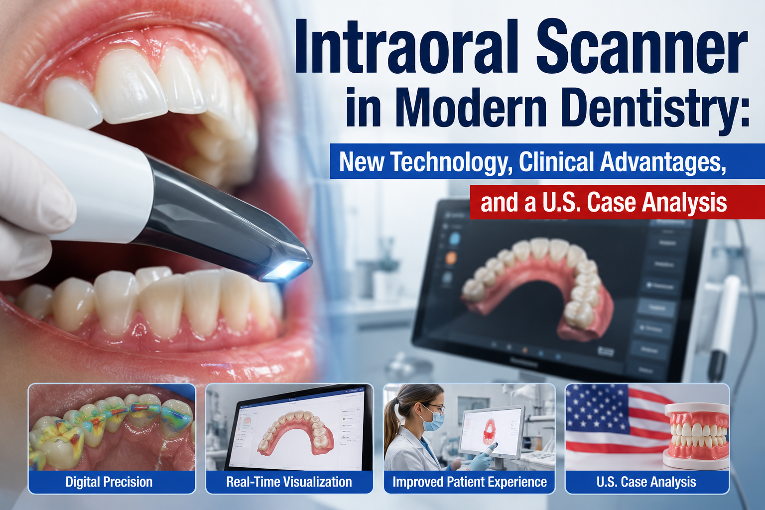
As a dentist working with modern digital dentistry in the United States and following developments in both American and German dental technology, I have seen intraoral scanning move from an emerging technology to an increasingly practical part of everyday dental care.
An intraoral scanner (IOS) is a handheld digital device that captures optical information from inside the patient's mouth and converts it into a three-dimensional digital model. Instead of using impression material and a traditional tray to record the teeth, the dentist can scan the oral structures directly.
The resulting digital file can be used for applications such as:
- Crowns
- Bridges
- Inlays and onlays
- Clear aligners
- Implant restorations
- Occlusal appliances
- Implant surgical guides
- Digital study models
- Treatment planning
The American Dental Association describes intraoral scanners as tools that can create digital impressions for crowns, inlays, onlays, fixed partial dentures, aligners, occlusal devices, and implant surgical guides.
From a clinical perspective, however, I would make one point very clear:
Digital does not automatically mean perfect.
The quality of an intraoral scan depends on the scanner, software, scanning technique, operator experience, moisture control, anatomy, restoration type, and clinical situation.
What Is an Intraoral Scanner?
An intraoral scanner is a small digital scanning device that the dentist moves around the patient's mouth.
The scanner captures many individual images or measurements and software combines them into a three-dimensional representation of the patient's teeth and surrounding oral structures.
Instead of producing a physical impression, the dentist obtains a digital impression.
The digital model can then be transferred electronically to compatible dental software, laboratories, CAD/CAM systems, or other digital workflows.
The American Dental Association notes that intraoral scanners can be used as standalone systems or integrated with technologies such as CBCT, 2D imaging, dental laboratory milling, and 3D printing.
This connectivity is one of the most important innovations.
Traditional Impression vs. Digital Scan
For decades, dentists commonly used impression trays filled with materials such as alginate or elastomeric impression materials.
The patient bites or holds the tray in position while the material sets.
For some patients, this experience can be uncomfortable.
Digital scanning changes the process.
Instead of:
Tray → Impression Material → Setting Time → Removal → Physical Model
the digital workflow can become:
Scanner → Digital 3D Model → CAD Design → Laboratory or 3D Printer
The process can be faster and more comfortable in many clinical situations. A systematic review found that intraoral scanning was generally faster than conventional impressions and was generally preferred by patients.
Why Is Intraoral Scanning Considered an Innovation?
The innovation is not simply replacing impression material with a camera.
The larger innovation is that dentistry can become a connected digital workflow.
A scan can potentially move through several stages:
Patient
↓
Intraoral Scan
↓
Digital Treatment Planning
↓
CAD Design
↓
Dental Laboratory
↓
Milling or 3D Printing
↓
Final Restoration
This can reduce the number of physical steps involved in some workflows.
The ADA describes digital dentistry as an expanding area involving intraoral scanning, imaging, artificial intelligence, electronic health records, and 3D printing.
Major Advantages of Intraoral Scanners
1. Improved Patient Comfort
One of the most obvious advantages is eliminating or reducing the need for conventional impression material.
Some patients have a strong gag reflex and find traditional impressions unpleasant.
With an intraoral scanner, the dentist uses a relatively small scanning wand rather than filling a large tray with impression material.
Research has found that patients generally prefer intraoral scanning over conventional impressions in many restorative situations.
2. Real-Time Visualization
This is one of the most useful advantages for both the dentist and patient.
As the dentist scans, the digital model appears on a computer screen.
I can potentially see:
- Tooth surfaces
- Preparation margins
- Missing areas
- Scan gaps
- Occlusal relationships
- Areas that need rescanning
Instead of waiting for a physical impression to be poured into a model, the dentist can evaluate the digital result immediately.
The ADA notes that digital scanning allows dentists to view digital files and use them for patient communication and treatment planning.
3. Selective Rescanning
Imagine taking a conventional impression and discovering a small defect.
Often, the impression may need to be repeated.
With digital scanning, the dentist can frequently identify the problematic area on the screen and rescan that region.
This can reduce unnecessary repetition.
However, the dentist must still verify that the final scan is clinically acceptable.
4. Faster Communication With the Dental Laboratory
Digital files can be transferred electronically.
Instead of physically shipping an impression or stone model, the dental laboratory can receive the digital case.
This can potentially reduce delays in communication.
The ADA has described digital workflows as a way to streamline communication between dental practices and laboratories.
5. Digital Storage
A conventional impression is a physical object.
A digital scan is a digital file.
That can make long-term storage, retrieval, and duplication easier within an appropriately managed digital dental record system.
A digital model can also potentially be compared with later scans.
6. Monitoring Tooth Wear
This is an especially interesting emerging application.
A patient's teeth can be digitally scanned at different points in time.
The dentist may compare the models to evaluate changes in:
- Tooth wear
- Tooth position
- Gingival contours
- Restoration shape
- Occlusion
Modern intraoral scanning systems and software are increasingly capable of supporting longitudinal digital workflows.
A recent systematic review notes that newer IOS technologies and software have expanded beyond impressions into applications such as monitoring tooth wear and soft-tissue changes.
7. Orthodontic Applications
Intraoral scanners have become particularly useful in orthodontics.
Digital scans can be used for:
- Treatment planning
- Digital models
- Clear aligner workflows
- Monitoring tooth movement
- Retainer fabrication
A systematic review of intraoral scanning in orthodontics found satisfactory to excellent reproducibility and reported advantages in scanning time and patient comfort, although accuracy varies according to the system and clinical circumstances.
8. Implant Dentistry
Intraoral scanning can also be incorporated into implant workflows.
For example, the dentist may capture:
- Implant position
- Adjacent teeth
- Soft tissues
- Occlusion
- Scan body information
The digital scan can then be combined with other digital information for prosthetic or surgical planning.
However, implant scanning has unique technical challenges.
A 2024 systematic review found that multiple factors can affect implant-scan accuracy, including scan-body characteristics and scanning conditions.
Therefore, an implant scan should never be considered automatically accurate simply because it is digital.
9. Integration With CAD/CAM
One of the biggest advantages is the connection between scanning and computer-aided design and manufacturing.
A typical workflow might look like:
Scan
→
Digital preparation
→
CAD design
→
Manufacturing
→
Restoration
Depending on the practice and laboratory, manufacturing may involve milling or additive manufacturing such as 3D printing.
This creates the possibility of a highly integrated digital dental workflow.
10. Better Patient Education
Patients often understand dentistry better when they can see their own teeth on a screen.
Imagine a dentist showing a patient:
"Here is the tooth before treatment."
Then:
"Here is the digital preparation."
Then:
"Here is the proposed crown."
This visual communication can make complex dental treatment easier to understand.
The ADA specifically notes that digital files can help dentists show patients their scans and visualize treatment plans.
U.S. Case Analysis: Digital Crown Workflow
Patient
Location: New York, USA
Age: 56
Chief concern: Broken lower molar
Treatment under consideration: Full-coverage crown
The patient presents with a fractured posterior tooth.
After clinical examination, the dentist determines that a crown may be appropriate.
The patient has a strong gag reflex and reports previous difficulty tolerating conventional impressions.
This is a situation in which an intraoral scanner may offer a significant practical advantage.
Step 1: Clinical Examination
Before scanning, I would evaluate:
- Remaining tooth structure
- Periodontal health
- Tooth mobility
- Pulpal status when indicated
- Occlusion
- Adjacent teeth
- Opposing tooth
- Existing restorations
The scanner does not replace the examination.
It supplements it.
Step 2: Tooth Preparation
The tooth is prepared according to the selected restorative plan.
The dentist must ensure that the preparation is clinically appropriate before scanning.
This is important because:
A highly accurate scan of a poorly prepared tooth is still a poor starting point for a restoration.
Digital technology cannot correct an incorrect clinical preparation.
Step 3: Digital Scanning
The dentist scans:
- Prepared tooth
- Adjacent teeth
- Opposing arch
- Bite relationship
The scanner produces a digital 3D model.
The dentist can immediately inspect the screen.
If an area is missing or distorted, it can be rescanned.
Step 4: Digital Laboratory Communication
The completed digital scan can be transferred to the dental laboratory.
The laboratory can use the digital information to design the restoration.
The digital workflow can reduce physical impression handling and may improve communication efficiency.
The ADA reports that digital scanning can streamline impression-making and reduce chair time and retakes in appropriate workflows.
Step 5: CAD Design
The laboratory digitally designs the crown.
The software can help establish:
- Tooth anatomy
- Contacts
- Occlusion
- Margins
- Emergence profile
- Overall restoration dimensions
The dentist and laboratory may communicate digitally about the case.
Step 6: Manufacturing
The restoration can then be fabricated using an appropriate manufacturing workflow.
Depending on the practice and laboratory, this may involve:
- Milling
- 3D printing for certain components
- Laboratory fabrication
- Hybrid digital/analog techniques
Step 7: Clinical Try-In
The dentist evaluates:
- Marginal fit
- Contacts
- Occlusion
- Esthetics
- Function
- Patient comfort
Even with an excellent digital scan, the final restoration still requires clinical verification.
Case Analysis: Why the Technology Helped
In this hypothetical case, the intraoral scanner provided several advantages:
Patient comfort
No large conventional impression tray was required.
Immediate feedback
The dentist could see the scan immediately.
Selective rescanning
A questionable region could be rescanned.
Digital laboratory communication
The scan could be transferred electronically.
Digital documentation
The digital model could become part of the patient's digital workflow.
Efficient workflow
The impression process could be streamlined.
This illustrates the real value of intraoral scanning:
The scanner is not simply a replacement for impression material. It can become the entry point into an entire digital restorative workflow.
How Accurate Are Intraoral Scanners?
This is where dentists need to be careful.
The answer is:
They can be highly accurate, but accuracy depends on the clinical situation.
Research has found that intraoral scanners can provide clinically acceptable digital impressions in many applications. However, different scanner systems and scanning conditions produce different results.
For many tooth-supported restorations and shorter-span cases, digital impressions can be highly effective.
However, complete-arch scans and certain edentulous or complex cases can present greater challenges.
The ADA specifically notes that while intraoral scanners can provide accurate impressions for tooth- and implant-supported crowns and short-span fixed prostheses, complete-arch scans and some removable prosthodontic applications may be less predictable than conventional methods.
What Can Reduce Scanning Accuracy?
Several factors can affect scan quality.
Saliva
Excess moisture can interfere with optical scanning.
Blood
Bleeding around preparation margins can make tissue and margins difficult to capture.
Deep subgingival margins
Margins located deep beneath the gingiva can be challenging.
Limited access
Posterior teeth can be difficult to scan.
Operator technique
The scanning path and technique matter.
Scanner technology
Different systems use different hardware and software.
Long-span scanning
The potential for accumulated errors can become more significant as the scanned area increases.
Research has shown that scanning strategy and dry conditions can influence digital impression accuracy.
Digital Does Not Mean Error-Free
This is one of the most important messages I would give patients.
An intraoral scanner can produce an impressive-looking 3D model.
But a visually beautiful scan is not automatically a clinically accurate scan.
The dentist must evaluate:
- Completeness
- Margins
- Occlusion
- Surface quality
- Distortions
- Stitching errors
- Missing areas
Operator training remains important.
The ADA has noted that dentists using intraoral scanners may need training to manage variables that can reduce scanning accuracy.
Intraoral Scanner Advantages vs. Traditional Impressions
| Feature | Intraoral Scanner | Conventional Impression |
|---|---|---|
| Patient comfort | Often excellent | Can be uncomfortable |
| Digital workflow | Yes | Usually requires additional digitization |
| Immediate visualization | Yes | No |
| Selective rescanning | Yes | Usually repeat impression |
| Laboratory transmission | Digital | Physical shipment or scanning |
| Physical impression storage | No | Yes |
| Moisture sensitivity | Yes | Also clinically relevant |
| Operator learning curve | Yes | Also requires skill |
| Complete-arch accuracy | Can be challenging | May be advantageous in some situations |
| Patient education | Excellent | Limited |
| CAD/CAM integration | Excellent | Requires additional steps |
The key is not that digital scanning is universally superior.
Rather:
Different clinical situations may favor different impression techniques.
New Technology: AI-Assisted Scanning
One of the most interesting developments is the integration of artificial intelligence and advanced software into digital scanning.
Potential capabilities include:
- Automatic identification of teeth
- Detection of missing scan areas
- Improved image processing
- Noise reduction
- Soft-tissue management
- Automated digital model analysis
- Tooth-wear monitoring
- Treatment visualization
A recent review describes newer IOS software as increasingly capable of supporting additional applications beyond traditional digital impressions.
However, AI should be considered a clinical support technology, not an independent dentist.
The clinician remains responsible for diagnosis and treatment decisions.
New Technology: Digital Twins of the Patient
Another exciting concept is the development of increasingly comprehensive digital representations of a patient's oral condition.
A future digital workflow may combine:
Intraoral scan
CBCT
Digital photographs
Radiographs
Facial scanning
Treatment history
to create a highly detailed digital representation of the patient's oral anatomy.
The goal is not simply to create a 3D picture.
The goal is to create a connected digital environment for:
- Diagnosis
- Treatment planning
- Simulation
- Manufacturing
- Monitoring
- Patient communication
This is one of the directions in which digital dentistry is evolving.
Germany and the Development of Digital Dentistry
Germany has played an important role in the development of CAD/CAM and digital dental manufacturing.
From a European clinical perspective, digital scanning is particularly valuable when it is integrated with:
- CAD/CAM
- Digital laboratory workflows
- Milling
- 3D printing
- Implant planning
- Digital orthodontics
The underlying principle is the same in Germany and the United States:
Capture accurate clinical data → process it digitally → design the treatment → manufacture the restoration → clinically verify the result.
The technology may differ between practices, but the clinical fundamentals remain the same.
Frequently Asked Questions
What is an intraoral scanner?
An intraoral scanner is a handheld digital device that captures the patient's oral structures and creates a three-dimensional digital model.
Is an intraoral scan better than a traditional impression?
Not in every situation.
Digital scanning offers important advantages in comfort, workflow, visualization, and communication, but conventional impressions can still be useful or preferable for certain complex cases.
Does an intraoral scanner hurt?
Generally, no. The scanner is moved around the mouth without impression material setting around the teeth. Some patients may still experience mild discomfort from cheek retraction or the scanner head.
Is an intraoral scanner safe?
Intraoral scanners are designed to be used in the mouth according to their manufacturer's instructions. The specific device, infection-control procedures, and clinical workflow matter.
How long does an intraoral scan take?
Scanning time varies significantly by scanner, operator, arch size, clinical conditions, and workflow. Modern systems can make scanning relatively quick, but the dentist should prioritize scan quality over speed.
Can an intraoral scanner replace dental X-rays?
No.
An intraoral scan primarily captures surface geometry. Radiographs and CBCT can provide information about structures that optical scanning cannot visualize.
Can an intraoral scanner detect cavities?
Some modern systems and software may assist with identifying certain abnormalities, but an intraoral scan should not automatically be considered a substitute for a comprehensive caries examination or appropriate radiographs.
Can an intraoral scanner be used for dental implants?
Yes. Digital scans can be incorporated into implant restorative and planning workflows. However, implant scanning has technical challenges, and scan accuracy depends on factors including scan-body design, scanning technique, and clinical conditions.
Can an intraoral scanner be used for crowns?
Yes. Crowns are one of the established applications for digital impressions.
Can an intraoral scanner be used for veneers?
Yes. Digital scans can support cosmetic and restorative workflows, although photographs, shade analysis, clinical examination, and other records may also be necessary.
Can an intraoral scanner be used for Invisalign or clear aligners?
Digital scans can be used for orthodontic treatment planning and aligner fabrication workflows.
Does the scanner replace the dentist?
Absolutely not.
The scanner captures information. The dentist interprets that information and determines the diagnosis and treatment plan.
Are digital impressions more comfortable?
Evidence generally indicates that patients tend to prefer intraoral scanning over conventional impression procedures, particularly because it avoids some of the discomfort associated with impression materials and trays.
Are digital scans always more accurate?
No.
Accuracy varies by scanner, technique, clinical situation, scan length, moisture, anatomy, and other factors. Some complex complete-arch and edentulous applications remain challenging.
Can my dentist show me the scan?
Often, yes. Many systems display the digital model on a monitor, allowing the dentist to explain findings and demonstrate treatment options.
Can an old digital scan be compared with a new scan?
Potentially, yes. Digital models can support longitudinal comparison, including monitoring changes in tooth position and tooth wear, depending on the software and workflow.
Advantages for the Patient
From a patient's perspective, the biggest advantages may be:
More comfortable impressions
No large tray filled with setting material in many cases.
Better communication
You can see your own teeth in 3D.
Faster digital communication
The laboratory can receive digital information electronically.
Easier treatment visualization
The dentist can potentially show proposed restorations or orthodontic changes digitally.
Digital records
The scan can become part of the patient's digital documentation.
Potentially fewer retakes
When a scan contains an incomplete area, the dentist can often rescan the specific region rather than repeating an entire conventional impression.
Advantages for the Dentist
For the dental professional, intraoral scanning can provide:
- Faster workflows
- Immediate visualization
- Digital storage
- Easier laboratory communication
- Selective rescanning
- CAD/CAM integration
- Improved patient education
- Digital treatment planning
- Integration with 3D printing
- Potential longitudinal monitoring
The ADA reports that dentists using intraoral scanners have identified improved efficiency and clinical outcomes among the major reasons for adopting the technology.
My Clinical Perspective
As a dentist evaluating digital dentistry from both the U.S. and German technology perspectives, I see intraoral scanning as one of the most important bridges between traditional clinical dentistry and fully digital workflows.
But I would never recommend choosing a scanner simply because it is the newest model.
The important questions are:
Does it provide clinically appropriate accuracy?
Does the dentist know how to use it properly?
Can the practice maintain good moisture control and scanning technique?
Does the laboratory have a compatible digital workflow?
Is the technology appropriate for the patient's specific treatment?
Those questions matter more than the marketing label on the scanner.
Final Conclusion
Intraoral scanners represent a major innovation in modern dentistry.
They can transform the impression process from a physical, sometimes uncomfortable procedure into a digital workflow that provides immediate visualization and connects the dental office with laboratories and CAD/CAM manufacturing.
Evidence supports important benefits such as patient comfort, efficiency, and clinically useful accuracy in many applications, while also showing that limitations remain in certain complex situations.
The future is likely to involve even greater integration between:
Intraoral Scanning
→ AI
→ Digital Imaging
→ CAD/CAM
→ 3D Printing
→ Digital Treatment Planning
→ Long-Term Monitoring
But technology should always serve the clinical goal.
The best digital dentistry is not simply the dentistry with the most advanced equipment.
It is the dentistry that uses technology appropriately, accurately, and thoughtfully to improve diagnosis, communication, comfort, efficiency, and patient care.
Related Clinical Cases
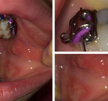
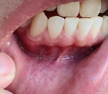
Patients with lower front gum inflammation may also experience tartar buildup, periodontal disease, gum abscesses, or tooth decay. Explore these related clinical case studies.


Other topics:
Other Dental Clinic