


Cheap Dental Implants Europe vs Philippines: Why Retirees Can Save Thousands

Topics:
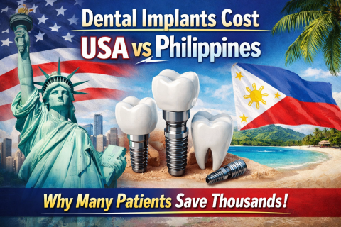
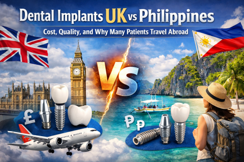
Dental implants are widely considered the best long-term solution for replacing missing teeth. They look natural, function like real teeth, and can last for decades with proper care. However, one major challenge for many patients—especially retirees—is the high cost of dental implants in Europe.
Across Europe, dental implant procedures can cost thousands of euros per tooth. Because of this, many patients are now exploring affordable options abroad. One destination gaining strong attention is the Philippines.