Broken Back Tooth with Deep Cavity Case Analysis
Severity:
Teeth Problems:
Dental Case Analysis: Broken Back Tooth with Deep Cavity
Case Overview
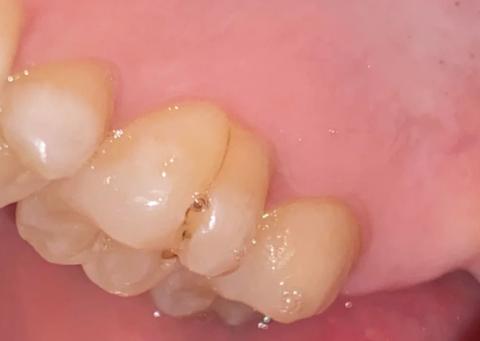
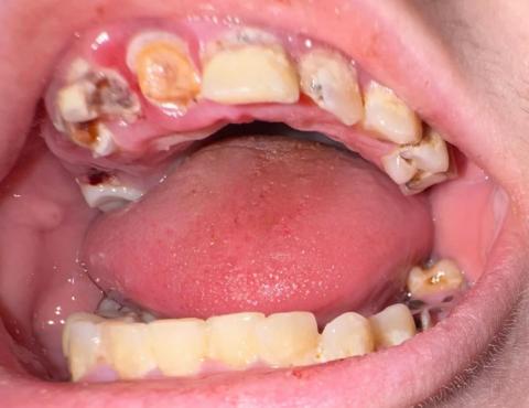
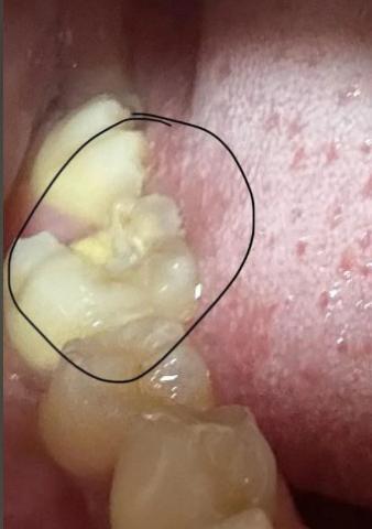
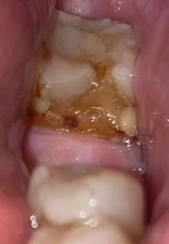
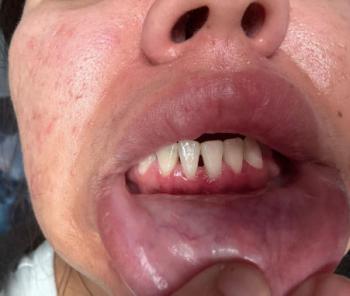
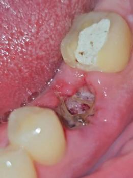
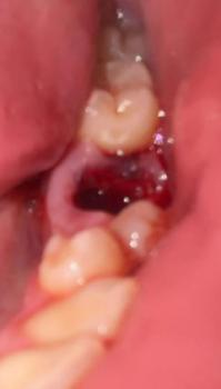
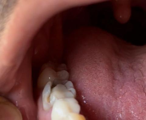
The image shows a posterior tooth located near the tongue side with visible enamel breakdown and a deep cavity. The tooth structure appears weakened, suggesting advanced decay and possible fracture.
Zoom 100% Visual Examination
Observed Findings
-
Back tooth with broken enamel edge
-
Deep cavity extending into dentin