From Pain to Perfect — We Care for Your Smile.

Root Problems: Root Canal Anatomy - A USA Dentist’s Zoom-In Clinical Review and Case Analysis
External root resorption is a relatively uncommon but important dental condition in which the body progressively removes part of the outer surface of a tooth root. Unlike ordinary tooth decay, the damage occurs along the root structure and can remain difficult to detect during the early stages.
From a California dental perspective, the key clinical challenge is determining why the resorption is occurring, how far it has progressed, whether the tooth can still be preserved, and whether the process is active or has become inactive.
This case-study style review explains how a USA dental team may evaluate external root resorption, what imaging can reveal, possible treatment approaches, and the questions patients should ask before deciding whether to save or replace the affected tooth.
USA Case Study: Progressive Root Damage
Patient profile: Adult patient presenting to a California dental clinic with intermittent discomfort involving an upper front tooth.
The patient did not initially report severe pain. A routine dental examination identified an unusual contour around the root on radiographic imaging.
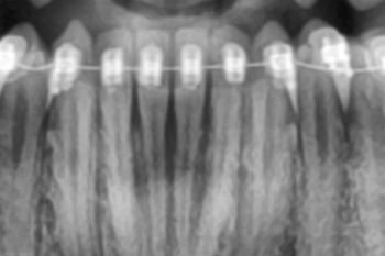
A conventional periapical X-ray suggested irregular loss of root structure. Because two-dimensional imaging could not completely establish the location and extent of the defect, the dentist recommended 3D CBCT imaging.
The CBCT examination demonstrated an area of external root surface destruction that was localized rather than representing ordinary internal decay.
Clinical Question #1: Is the tooth still restorable?
The first priority is not automatically extraction.
The dental team evaluates:
-
Remaining root length
-
Location of the resorption
-
Amount of supporting bone
-
Tooth mobility
-
Pulpal status
-
Periodontal condition
-
Presence of infection
-
Whether the resorption is active
-
Whether restoration or surgical access is possible
A tooth with limited, accessible external resorption may have a substantially different prognosis from a tooth with extensive circumferential root destruction.
What Causes External Root Resorption?
External root resorption can have several potential associations, including:
-
Dental trauma
-
Previous orthodontic treatment
-
Pressure from adjacent structures
-
Impacted teeth
-
Certain inflammatory processes
-
Periodontal or periapical inflammation
-
Previous dental procedures
-
Idiopathic cases where no clear cause is identified
Importantly, the cause cannot always be determined from an X-ray alone.
External vs. Internal Root Resorption
One of the most important diagnostic distinctions is whether the resorption originates externally or internally.
External root resorption begins from the external root surface.
Internal resorption originates within the tooth and expands outward from the internal root canal space.
Because treatment can differ considerably, accurately identifying the origin and location of the lesion is critical.
Clinical Review: Why CBCT Can Matter
A conventional dental X-ray provides a two-dimensional representation of a three-dimensional structure.
CBCT can provide additional information about:
-
Exact lesion location
-
Circumferential root involvement
-
Remaining root thickness
-
Relationship to the cortical bone
-
Nearby anatomical structures
-
Surgical accessibility
CBCT is not automatically required for every dental patient. The dentist should determine whether the additional imaging information is justified for the clinical question being investigated.
Treatment Strategy
Treatment depends heavily on the type, location, activity, and severity of the resorption.
1. Monitor a Stable Lesion
If the process appears inactive and the tooth remains clinically healthy, the dentist may recommend observation with appropriate follow-up.
2. Remove the Resorptive Tissue
When the defect is accessible, treatment may involve surgical exposure and removal of resorptive tissue followed by repair of the root defect.
3. Endodontic Treatment
If the pulp has become involved or the clinical diagnosis indicates endodontic treatment, root canal therapy may become part of the treatment plan.
However, not every external resorption case automatically requires a root canal. The treatment must be based on the actual pulpal and periodontal findings.
4. Extraction and Tooth Replacement
When extensive root destruction makes the tooth unrestorable, extraction may be considered.
Replacement options can include:
-
Dental implant
-
Fixed dental bridge
-
Removable partial denture
-
Other individualized restorative solutions
If an implant is being considered, the dentist must also evaluate the remaining bone and whether bone preservation or augmentation may be necessary.
California Dental Expert Review
From a USA clinical perspective, one of the biggest mistakes is treating the X-ray image rather than treating the entire biological problem.
The dentist should establish:
What caused the resorption?
Is it active?
Where exactly is it located?
How much root remains?
Is the tooth structurally restorable?
Is the pulp healthy?
Is there periodontal involvement?
Only after answering these questions should the treatment plan be finalized.
Patient Review: What Symptoms Should You Watch For?
External root resorption can sometimes progress with few obvious symptoms.
Possible findings may include:
-
Tooth discoloration
-
Mild or intermittent discomfort
-
Tooth mobility
-
Localized gum swelling
-
Sensitivity
-
Changes detected during routine X-rays
-
Unexpected changes following previous dental trauma or orthodontic treatment
Some patients may have no symptoms at all.
That is why an apparently normal tooth should not automatically be assumed to have a normal root structure.
Red Flags Requiring Prompt Dental Evaluation
Seek prompt professional dental evaluation if you develop:
-
New swelling around a tooth
-
Increasing pain
-
Tooth mobility
-
Significant discoloration
-
Drainage or pus
-
Persistent sensitivity
-
Facial swelling
-
Fever or systemic illness associated with dental symptoms
Facial swelling, difficulty breathing or swallowing, or rapidly spreading swelling can represent a dental emergency and requires urgent medical evaluation.
Case Outcome Framework
In a real California dental case, the final outcome would depend on the individual patient's examination and imaging.
A favorable scenario may involve:
Early detection → accurate 3D diagnosis → removal/control of the resorptive process → restoration → long-term monitoring.
A more advanced scenario may involve:
Extensive root destruction → poor structural prognosis → extraction → bone preservation/augmentation when appropriate → future tooth replacement.
The objective is not simply to remove a damaged tooth. When possible, the objective is to determine whether the natural tooth can be predictably preserved.
Questions to Ask Your Dentist
Before treatment, patients can ask:
-
Is this definitely external root resorption?
-
How extensive is the resorption?
-
Is the lesion active?
-
What caused it?
-
Does the pulp remain healthy?
-
Do I need CBCT imaging?
-
Can the tooth be restored?
-
What is the expected prognosis if we try to save it?
-
What happens if the tooth is extracted?
-
If an implant is recommended, will bone grafting be necessary?
Dental Hub Takeaway
External root resorption is a root problem that deserves careful diagnosis rather than a one-size-fits-all treatment.
Early identification can sometimes provide an opportunity to preserve the natural tooth. Advanced resorption, however, may compromise the structural integrity of the root and require extraction and replacement.
A California dental specialist, endodontist, periodontist, or restorative dentist may be involved depending on the location and complexity of the lesion.
Important: This case study is educational and does not diagnose an individual patient. External root resorption requires an in-person dental examination and appropriate imaging before treatment decisions are made.
Related Clinical Cases
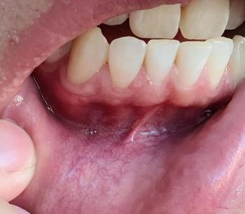
Patients with lower front gum inflammation may also experience tartar buildup, periodontal disease, gum abscesses, or tooth decay. Explore these related clinical case studies.


Other topics:
Other Dental Clinic