Multiple Tooth Decay and Plaque Buildup Case Evaluation
Severity:
Teeth Problems:
Multiple Tooth Decay and Plaque Buildup Case Evaluation
Case Overview
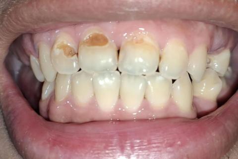
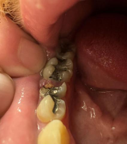
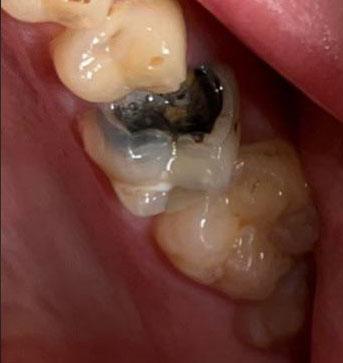
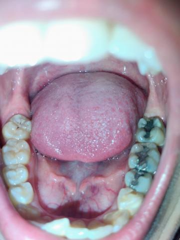
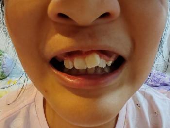
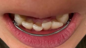
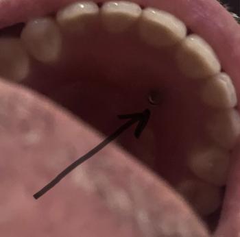
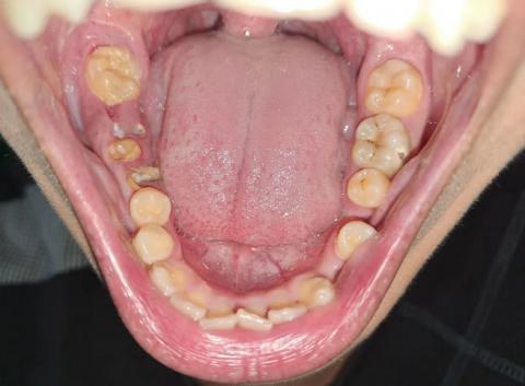
The image shows a full lower arch view with visible multiple carious lesions, plaque accumulation, and structural enamel damage. Several posterior teeth present brown cavitated areas indicating active dental decay. The lower anterior teeth also show crowding and tartar buildup.
This condition appears progressive and requires immediate dental evaluation to prevent further complications.