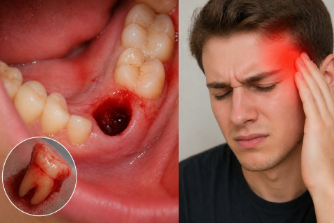
Headache After Tooth Extraction: Causes, Timeline, and When to Worry
Severity:
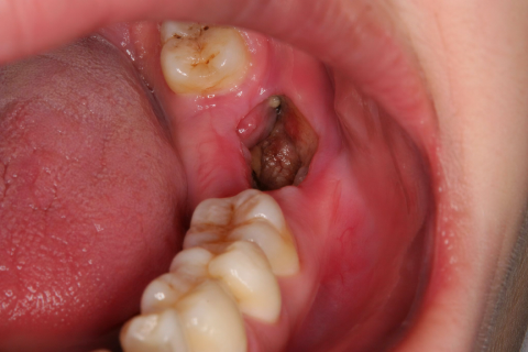
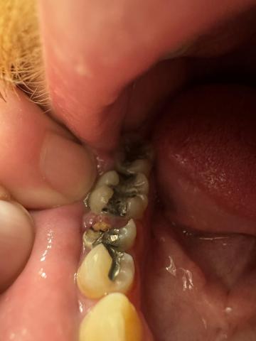
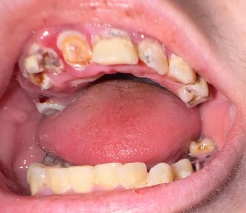
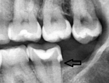
Teeth Problems:
A Clinical Guide from an Illinois Dental Bone Graft Expert (10 Years Experience)
Headaches after a tooth extraction are more common than most patients expect—and often more confusing than painful.
Many people assume something is wrong with their brain or sinuses. In reality, after 10 years of treating extraction and implant patients in Illinois, I can tell you:
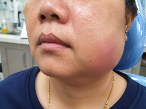
Most post-extraction headaches are not dangerous—they’re referred pain from your jaw and surrounding structures.