Bone Loss After Tooth Extraction in Delaware, USA: Expert Bone Graft Guide From a 25-Year Specialist
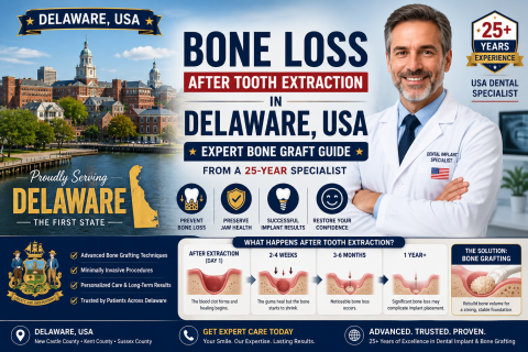
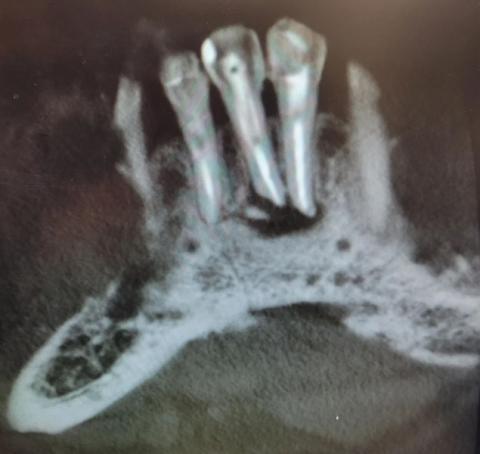
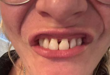
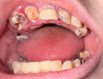
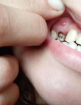
Tooth extraction is sometimes necessary to protect your oral health, but many patients in Delaware are surprised to learn that losing a tooth can also lead to bone loss in the jaw. This process may begin faster than expected, especially during the first few months after an extraction. Without proper care, bone shrinkage can affect facial appearance, chewing ability, speech, and future dental implant options.