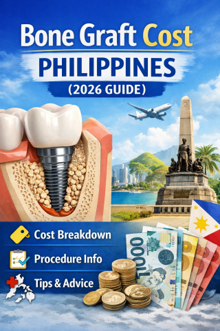


The Hard Truth About Bone Grafts in the Philippines Before Dental Implants

Topics:
Most Filipino patients want dental implants.
Very few are ready for what comes before implants.
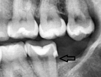
If you lost a tooth years ago, your jawbone has already shrunk. No bone = no implant. And that’s where bone grafting comes in.
Let’s break this down properly.
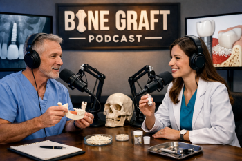
PART A: The Graft Guide (The “What”)
Bone grafting is not optional when you have moderate to severe bone loss. It is biological reconstruction.
Below is a straightforward guide to the graft materials commonly available in the Philippines.