Clear Orthodontic Braces With Active Tooth Alignment – Dental Case Analysis
Severity:
Teeth Problems:
Dental Case Analysis – Clear Orthodontic Braces With Active Tooth Alignment
Case Overview
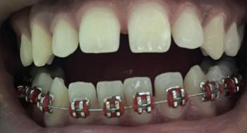
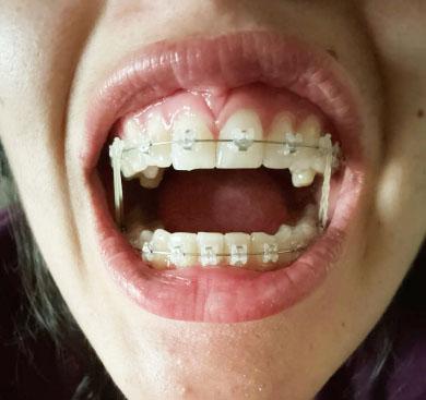
This image shows a patient undergoing active orthodontic treatment using clear or ceramic braces on both the upper and lower teeth. Teeth are in an alignment and leveling phase, with visible archwires and elastic ligatures. The surrounding soft tissue appears generally healthy, with mild tension expected during tooth movement.