Severe

Large Red Gum Swelling Near Tooth - Dental Case Analysis

Severity: 

Dental Case Analysis – Large Red Gum Swelling Near Posterior Tooth

Case Overview

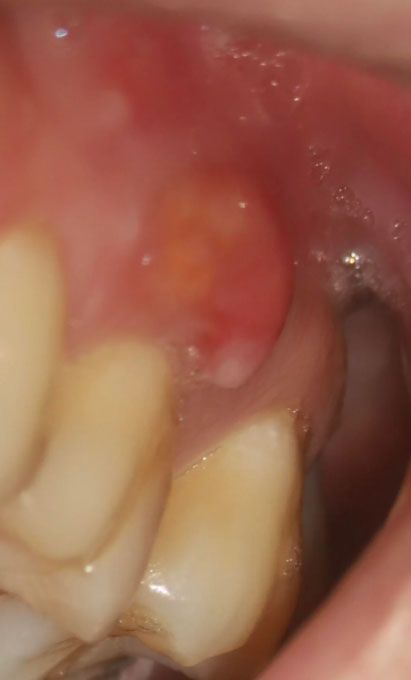
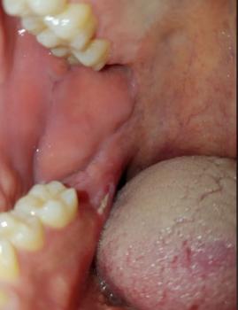
This intra-oral image shows a large, red, swollen lesion on the gum tissue adjacent to a posterior tooth. The appearance is consistent with an active localized gum infection, likely involving deeper periodontal tissues and possibly the underlying bone if not treated promptly.


Full Analysis and Diagnosis

Clinical Findings

  • Prominent red, dome-shaped swelling on gingiva

  • Inflamed, shiny soft tissue

Facial Skin Infection Possibly Linked to Dental Cause - Case Analysis

Severity: 

Dental Case Analysis – Facial Skin Infection Possibly Related to Dental Source

Case Overview

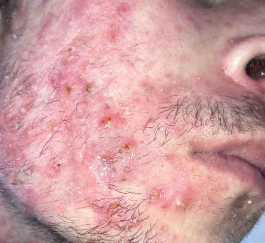
This image shows a diffuse inflammatory facial rash involving the lower cheek, beard area, and jawline. Multiple inflamed papules, pustules, and crusted lesions are present. The distribution closely follows the mandibular region, raising concern for a possible underlying dental or jaw-related inflammatory source.

Recurrent Jawbone Infection With Facial Rash - Dental Case Analysis

Severity: 

Dental Case Analysis – Recurrent Jawbone Infection With Persistent Facial Rash

Case Overview

This case involves a patient with a history of bacterial infection in the lower jawbone affecting teeth 25 and 26, complicated by bone loss and incomplete root canal treatment due to canal calcification. Years later, imaging shows recurrent infection, alongside a persistent facial rash localized to the same area, causing emotional and physical distress.

Lower Molars on Both Sides With Orthodontic Appliances Case Analysis

Severity: 
Teeth Problems: 

What Is Seen in This Case

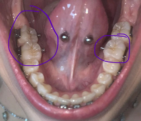
The image shows the lower jaw with molars on both the left and right sides, highlighted by circles. There are orthodontic components (brackets or attachments) present near the molars, and the chewing surfaces show plaque accumulation and early staining. The tongue piercings are visible but are not the main concern in this case.

This setup makes cleaning more difficult and increases the risk of decay around the molars.

Post Root Canal Infection With Sutures Case Analysis and Treatment Guide

Severity: 

What Is Seen in This Case

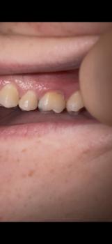
The image shows a treated tooth with sutures placed on the surrounding gum tissue. The area appears reddened and swollen, indicating recent root canal–related surgical treatment or management of an infection at the root area. The presence of sutures suggests that the infection required surgical access, drainage, or gum repair.

This is a normal appearance shortly after treatment but requires close monitoring.

Pages