Panoramic Dental X-Ray Case Analysis After Dental Treatment
Severity:
Teeth Problems:
What Is Seen in This X-Ray
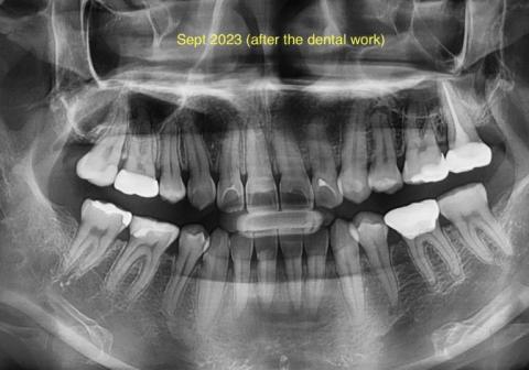
This panoramic dental X-ray (taken after dental work in September 2023) shows multiple restored teeth, including fillings and treated posterior teeth. The roots and surrounding bone are visible across the upper and lower jaws, allowing evaluation of healing, hidden infection, and overall oral health.
Some areas show treated teeth with restorations, while other regions require close monitoring for possible residual or recurrent infection.