Healthy Teeth, Happy Life.

Hidden Tooth Infections: Chronic Tooth Infection and Root Canal Infection
image hero:
A Canadian Dental Case Study and Clinical Analysis
Dental Hub Clinical Feature
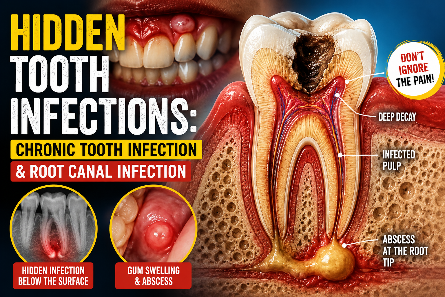
A tooth can look relatively normal on the outside while a significant infection is developing inside the root canal system or around the tip of the root. This is one reason chronic dental infections can be difficult for patients to recognize.
From a Canadian dental perspective, the key clinical question is not simply, “Does the tooth hurt?” The more important questions are: Is the dental pulp alive and healthy? Is there evidence of infection around the root? Is the tooth restorable? And what does the clinical examination and dental imaging reveal?
The Canadian Dental Association explains that bacteria can enter the pulp through deep decay, cracks or defective fillings. When the pulp becomes infected, an abscess can develop, sometimes producing pain or swelling and sometimes being discovered primarily through examination or radiographic imaging.
Case Study: The Tooth That “Didn’t Hurt”
Patient presentation
Consider a hypothetical Canadian patient in their late 40s who visits a dental clinic for a routine examination.
The patient reports:
-
No significant toothache
-
Occasional pressure when chewing
-
Mild bad taste that comes and goes
-
Previous large filling on a lower molar
-
No obvious facial swelling
-
No recent dental emergency
The patient assumes the tooth is healthy because there is little or no pain.
During examination, however, the dentist notices that the molar has extensive previous restorative work. A percussion test produces mild tenderness, and radiographic examination reveals a dark area around the root tip.
The clinical picture changes immediately.
The problem may not be a simple filling issue. The findings raise concern for pulpal disease and infection involving the tissues surrounding the root.
Clinical lesson
Absence of severe pain does not prove that a tooth is healthy.
In some chronic infections, drainage through a small opening in the gum can reduce pressure and therefore reduce pain. Canadian clinical guidance describes situations in which chronic suppuration can produce a gum boil or draining lesion while pain may be limited.
This is one of the most important concepts behind the term “hidden tooth infection.”
How a Hidden Root Canal Infection Develops
A typical pathway can look like this:
Dental decay or fracture
↓
Bacteria penetrate deeper tooth structure
↓
Dental pulp becomes inflamed
↓
Pulp damage becomes irreversible
↓
Pulp tissue dies
↓
Bacteria and inflammatory products extend through the root canal
↓
Inflammation develops around the root
↓
Bone destruction may become visible on dental imaging
↓
Chronic apical infection or abscess develops
The Canadian Dental Association notes that deep cavities, cracks and defective fillings can allow bacteria to enter the tooth. Root canal treatment removes infected, injured or dead pulp, followed by cleaning, disinfection, filling and sealing of the canal system.
Why Chronic Tooth Infection Can Be Difficult to Detect
1. The nerve may already be dead
A common misconception is:
“If the tooth is infected, it must hurt.”
Not necessarily.
When the dental pulp becomes severely damaged or dies, the sensory response can change dramatically. A tooth that previously reacted strongly to hot or cold may eventually stop responding.
That can create a dangerous false sense of security.
The patient may interpret the disappearance of pain as healing when the underlying disease process has actually progressed.
2. The infection may drain
A chronic infection can sometimes establish a drainage pathway through the gum.
The patient may notice:
-
A small pimple-like bump
-
Intermittent drainage
-
Bad taste
-
Occasional pus
-
Mild gum swelling
-
A recurrent area that appears and disappears
Because pressure is relieved through drainage, the tooth may not produce dramatic pain.
This is why a small “gum boil” should not simply be ignored.
3. The infection may only appear on imaging
A dentist may discover a periapical lesion during an examination even when the patient has minimal symptoms.
Radiographs can reveal changes around the root that are not visible by simply looking at the tooth.
This is particularly important when evaluating:
-
Previously root-treated teeth
-
Large restorations
-
Cracked teeth
-
Deep cavities
-
Discoloured teeth
-
Teeth with previous trauma
-
Teeth with unexplained tenderness
The Canadian Dental Association specifically notes that infection may be detected through dental X-rays or other changes involving the tooth.
Case Analysis: What Would the Dentist Investigate?
A thorough assessment does not depend on one symptom or one X-ray.
Step 1 — Medical and dental history
The dentist reviews:
-
Previous fillings
-
Previous root canal treatment
-
Trauma
-
Previous toothache
-
Sensitivity to hot and cold
-
Pain during chewing
-
Swelling
-
Drainage
-
Previous dental treatment
Step 2 — Visual examination
The dentist looks for:
-
Deep decay
-
Cracks
-
Discolouration
-
Defective restorations
-
Gum swelling
-
Sinus tracts
-
Tooth fractures
Step 3 — Pulp testing
Depending on the situation, the dentist may perform thermal or electrical pulp testing to determine whether the pulp is responsive.
The result must be interpreted alongside the other clinical findings.
Step 4 — Percussion and palpation
The dentist checks whether the tooth or surrounding tissues are tender.
Pain when tapping a tooth can indicate inflammation involving the periodontal ligament and tissues around the root.
Step 5 — Dental imaging
Radiographs help identify changes around the root.
Depending on the complexity of the case, additional imaging such as three-dimensional imaging may be considered by the treating clinician.
Root Canal Infection vs. Gum Infection
This distinction is critical.
A patient may experience swelling around a tooth, but the source can be different.
Endodontic infection
The primary problem originates within the tooth's pulp and root canal system.
Possible causes include:
-
Deep dental decay
-
Tooth fracture
-
Trauma
-
Failed previous restoration
-
Previous root canal complications
Periodontal infection
The primary disease involves the gum and supporting tissues surrounding the tooth.
Canadian public-health guidance describes periodontitis as a chronic inflammatory disease involving the tissues and bone supporting the teeth. Symptoms can include bleeding gums, persistent bad breath, loose teeth, pus or a bad taste and gum recession.
Why the distinction matters
A patient can have both endodontic and periodontal disease at the same time.
That makes diagnosis more complicated and may require coordinated periodontal and endodontic evaluation.
The Root Canal: What Is Actually Being Treated?
A root canal is not simply “filling a hole in the tooth.”
The objective is to remove diseased or infected pulp and eliminate microorganisms and contaminated tissue from the root canal system.
According to the Canadian Dental Association, treatment involves:
-
Local anesthetic
-
Isolation of the tooth
-
Access into the tooth
-
Removal of diseased pulp
-
Cleaning and enlargement of the canal system
-
Disinfection
-
Filling and sealing of the canals
-
Restoration of the tooth
The final restoration is also important.
A tooth that has undergone root canal treatment still requires protection from future decay and fracture. Depending on how much tooth structure remains and the location of the tooth, a filling or crown may be recommended.
Why Antibiotics Are Not the Main Treatment for Every Root Canal Infection
This is an important area where patients can become confused.
An antibiotic does not replace definitive treatment of an infected tooth.
Canadian dental guidance emphasizes that antibiotics should not routinely be prescribed for ordinary toothache or a localized dental abscess without systemic complications. Definitive dental treatment—such as root canal treatment, extraction and/or drainage—is generally required to address the source. Antibiotics may become appropriate when there are systemic complications or spreading infection, depending on the patient's circumstances.
Therefore:
Antibiotics may control bacterial spread in appropriate situations, but they do not mechanically clean and seal an infected root canal.
The infected source still needs dental treatment.
Case Study Progression
Imagine the patient delays treatment because the tooth is not painful.
Month 1
Minor chewing discomfort.
The patient ignores it.
Month 3
The tooth becomes less sensitive.
The patient assumes it has improved.
Month 6
A small gum bump develops.
It disappears after several days.
Month 9
Routine dental imaging reveals a periapical radiolucency.
The patient is surprised:
“How can I have an infection when I don't have a toothache?”
This is precisely why clinical examination matters.
The disease may have progressed silently.
What Happens If the Infection Is Ignored?
An untreated dental infection can remain localized, become chronic, or progress into surrounding tissues.
Potential consequences include:
-
Persistent inflammation
-
Bone destruction around the root
-
Recurrent abscess formation
-
Gum drainage
-
Increasing swelling
-
Tooth weakening
-
Tooth loss
-
Spread of infection into surrounding facial tissues
Health Canada notes that oral infections and abscesses can spread beyond the jaws.
A patient with facial swelling, rapidly progressing swelling, difficulty swallowing, difficulty breathing, significant systemic illness or difficulty opening the mouth should receive urgent medical and dental assessment.
When Is Root Canal Retreatment Necessary?
A tooth that has already received root canal treatment can occasionally develop persistent or recurrent disease.
Possible explanations may include:
-
Complex canal anatomy
-
Previously untreated canal anatomy
-
Persistent infection
-
Inadequate sealing
-
New decay
-
Fracture
-
Leakage around the restoration
The Canadian Dental Association notes that retreatment involves removing the previous canal filling material, cleaning and reshaping the canal system and refilling it. In selected cases, endodontic surgery may be considered when conventional treatment cannot adequately resolve the problem.
When Extraction Becomes the Better Option
Not every infected tooth can or should be saved.
The dentist evaluates whether the tooth is restorable.
Important considerations include:
-
Remaining tooth structure
-
Root fracture
-
Periodontal support
-
Bone loss
-
Canal anatomy
-
Previous treatment
-
Ability to restore the tooth
-
Overall prognosis
If a tooth cannot predictably be restored, extraction may become the appropriate treatment.
If the tooth can be successfully treated and restored, preserving the natural tooth may be preferable.
Canadian Patient Perspective
For patients in Canada, another important consideration is access to appropriate dental care and understanding what treatment is being proposed.
The Canadian Dental Care Plan includes endodontic services such as root canal treatment under its applicable eligibility and coverage rules. Certain procedures, including some retreatments and surgical procedures, may require preauthorization. Coverage depends on the plan's current criteria and the individual patient's circumstances.
Patients should confirm coverage and treatment eligibility directly with their dental provider and the applicable CDCP rules rather than assuming that every root canal or retreatment is covered in the same way.
The Most Important Clinical Lesson
A hidden tooth infection is not necessarily a mysterious disease.
Often, it is a disease that has become quiet.
The tooth may no longer produce dramatic pain because the pulp has been severely damaged or because chronic drainage has reduced pressure.
That is why dentists evaluate more than pain.
The diagnosis comes from the combination of:
History + clinical examination + pulp testing + periodontal evaluation + dental imaging + restorability assessment.
No single symptom should be used to diagnose a root canal infection.
Dental Hub Takeaway
If you have a tooth with:
-
A previous deep filling
-
A cracked or fractured tooth
-
Persistent chewing sensitivity
-
A recurring gum pimple
-
Bad taste or drainage
-
Tooth discolouration
-
Previous root canal treatment
-
Unexplained swelling
-
An abnormal dental X-ray
do not assume that the absence of severe pain means everything is normal.
A dentist—or an endodontist for a more complex case—can determine whether the problem originates inside the tooth, around the root, in the periodontal tissues, or from another condition.
The goal is not simply to eliminate pain. The goal is to identify and eliminate the source of disease while preserving the tooth when it can be predictably saved.
This article is educational and does not diagnose an individual patient. A dental examination and appropriate imaging are required to determine whether a specific tooth has a root canal infection.
Related Clinical Cases
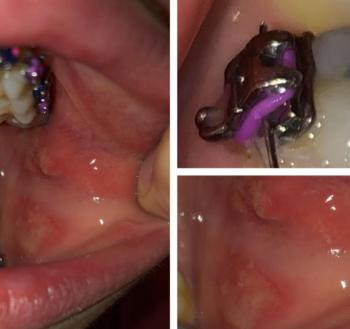
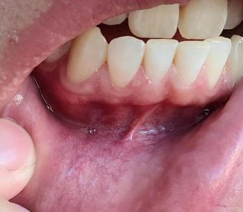
Patients with lower front gum inflammation may also experience tartar buildup, periodontal disease, gum abscesses, or tooth decay. Explore these related clinical case studies.



Other topics:
Other Dental Clinic