Is Dry Socket Dentist’s Fault? (Expert Insight from Osaka, Japan)
Topics:
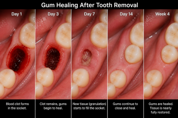
Dry socket is one of the most painful complications after a tooth extraction. Many patients immediately wonder: “Did my dentist do something wrong?”
The answer is not always straightforward.
As a dental specialist practicing in Osaka, I can confidently say that dry socket is rarely caused by dentist error alone. It is typically the result of multiple factors—many of which are beyond clinical control.