


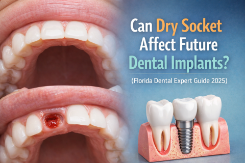
Can Dry Socket Affect Future Dental Implants? (Florida Dental Expert Guide 2025)
Topics:
Many patients worry that complications after tooth extraction—especially Dry Socket—might affect their chances of getting dental implants later. From the perspective of dental experts in Florida, the answer is reassuring:
Dry socket does not usually prevent future dental implants—but proper healing is essential.
What Is Dry Socket and Why It Matters
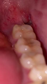
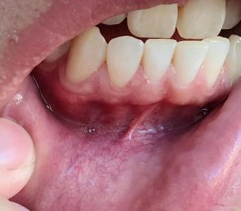
Dry Socket occurs when the protective blood clot is lost after extraction, exposing the bone and nerves.
Florida dental specialists explain: