We Bring Back the Sparkle in Your Smile.

Hidden Tooth Infections: Dental Abscess Analysis and Clinical Case Study
image hero:
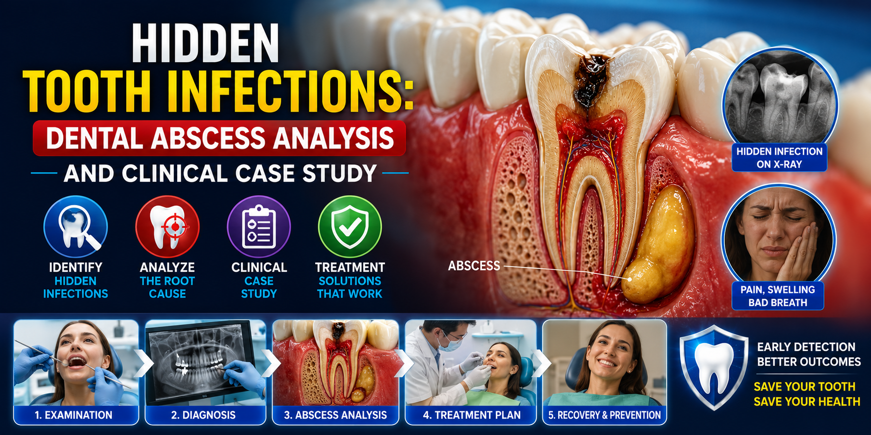
A tooth infection does not always announce itself with severe pain. In clinical practice, one of the most deceptive presentations is the hidden dental infection: a chronic infection that develops around the tooth root, beneath an existing restoration, inside a previously treated tooth, or within the surrounding bone while producing only mild or intermittent symptoms.
From a German dental clinical perspective, the central principle is straightforward: absence of pain does not prove absence of infection.
A patient may report that a tooth feels completely normal while radiographic examination reveals a periapical lesion, bone destruction, or evidence of chronic inflammation. These cases require careful diagnostic analysis rather than treatment based solely on symptoms.
1. Case Presentation
Patient: Adult patient
Primary complaint: Occasional pressure and mild discomfort in the posterior tooth region
Pain: Minimal and intermittent
Swelling: None externally visible
Fever: Absent
Medical history: No major systemic symptoms reported
Dental history: Previous large restoration on the suspected tooth
The patient initially considered the problem insignificant because there was no severe toothache.
During examination, however, the dentist identified subtle clinical findings:
-
Mild tenderness to percussion
-
Previous extensive restoration
-
Slight sensitivity during biting
-
Localized periodontal changes
-
No obvious facial swelling
-
No dramatic visible abnormality of the tooth
The clinical findings alone were not sufficient to establish the diagnosis.
Radiographic examination
A periapical radiograph demonstrated a radiolucent area around the root apex, suggesting inflammatory bone changes associated with the tooth.
Depending on the clinical situation, additional imaging such as CBCT may be considered when conventional radiographs do not adequately demonstrate the anatomy or extent of the lesion.
The important diagnostic question was therefore not simply:
"Does the tooth hurt?"
It was:
"Is the tooth biologically healthy, and what is happening around its root?"
2. Clinical Analysis: How a Tooth Infection Becomes Hidden
A chronic tooth infection can develop when bacteria gain access to the pulp through pathways such as:
-
Deep dental caries
-
Cracks or fractures
-
Failed restorations
-
Recurrent decay
-
Dental trauma
-
Previous pulp inflammation
-
Complications associated with root canal treatment
When the dental pulp becomes severely inflamed or necrotic, microorganisms and inflammatory products can extend through the root canal system toward the tissues surrounding the root.
The body's immune response then attempts to contain the infection.
This may produce a chronic inflammatory lesion around the root.
Interestingly, the patient may experience less pain as the pulp loses vitality. This creates one of the most important clinical misconceptions:
Less pain does not necessarily mean improvement.
In some cases, it may indicate that the pulp has become necrotic.
3. Dental Abscess vs. Chronic Apical Infection
The term dental abscess is commonly used broadly, but clinically several different conditions must be distinguished.
Acute dental abscess
An acute abscess may present with:
-
Significant toothache
-
Pain during chewing
-
Localized swelling
-
Tenderness
-
Pus formation
-
Possible fever or systemic illness
The patient usually recognizes that something is seriously wrong.
Chronic apical infection
A chronic infection may behave very differently.
Possible findings include:
-
Little or no spontaneous pain
-
Intermittent discomfort
-
Localized tenderness
-
A sinus tract or "gum boil"
-
Persistent unpleasant taste
-
Radiographic bone changes
-
Previous root canal treatment
-
Non-vital tooth
This is why chronic infections can remain undetected for considerable periods.
4. The "Gum Boil" That Should Not Be Ignored
One particularly important clinical clue is a small pimple-like swelling on the gum.
Patients sometimes assume this is simply a gum problem.
However, a localized sinus tract can represent drainage from an infection originating deeper around the tooth root.
The drainage may temporarily reduce pressure.
Consequently, the patient may experience:
Swelling → drainage → temporary relief → infection persists
The disappearance of swelling does not necessarily mean the infection has been eliminated.
The source of the infection must still be identified.
5. Case Study: The Pain-Free Infected Tooth
Initial presentation
A patient attended a dental examination without reporting significant pain.
The patient stated that the tooth had occasionally felt "different" but was still functional.
Clinical examination showed no major facial swelling.
Diagnostic investigation
The dentist performed:
-
Visual examination
-
Percussion testing
-
Palpation
-
Periodontal probing
-
Pulp vitality testing
-
Bite assessment
-
Periapical radiography
The radiograph revealed an area of bone loss around the root apex.
Diagnostic interpretation
The combination of:
-
Previous deep restoration
-
Abnormal pulp testing
-
Percussion sensitivity
-
Periapical radiographic changes
strongly suggested a disease process originating from the pulp and extending into the periapical tissues.
The important lesson is that clinical symptoms and radiographic findings must be interpreted together.
6. Why the Infection Can Remain Undetected
Several biological and anatomical factors can make an infection difficult to recognize.
1. Pulp necrosis
Once the pulp becomes necrotic, the tooth may no longer respond normally to temperature testing.
2. Chronic drainage
A sinus tract may provide an outlet for inflammatory fluid, preventing significant pressure buildup.
3. Slow progression
Some infections develop gradually rather than producing a sudden inflammatory episode.
4. Previous root canal treatment
A previously treated tooth can develop persistent or recurrent apical disease because of:
-
Missed canals
-
Inadequate cleaning
-
Persistent microorganisms
-
Leakage
-
New decay
-
Fracture
-
Complex canal anatomy
5. Individual pain response
Pain perception varies considerably between patients.
Therefore, symptoms alone cannot reliably exclude infection.
7. Diagnostic Strategy
A German dental clinician would generally approach a suspected chronic infection systematically rather than immediately assuming that every radiolucency represents an abscess.
Step 1: Patient history
The dentist asks about:
-
Previous toothache
-
Trauma
-
Root canal treatment
-
Previous swelling
-
Pain when biting
-
Temperature sensitivity
-
Previous dental procedures
-
Duration of symptoms
Step 2: Clinical examination
The dentist evaluates:
-
Tooth color
-
Restorations
-
Caries
-
Cracks
-
Gum condition
-
Sinus tracts
-
Local swelling
-
Periodontal probing
-
Percussion
-
Palpation
Step 3: Pulp testing
Appropriate vitality or sensibility testing can help determine whether the pulp is functioning.
Testing must be interpreted carefully and compared with neighboring control teeth.
Step 4: Radiographic assessment
Periapical radiography can identify:
-
Periapical radiolucency
-
Bone changes
-
Root morphology
-
Previous root canal treatment
-
Restoration problems
Step 5: Advanced imaging when indicated
CBCT may provide three-dimensional information when conventional imaging is insufficient, particularly in complex endodontic cases.
However, CBCT should not automatically replace conventional radiographs. Imaging should be justified by the diagnostic question.
8. Treatment Analysis
Treatment depends on the source, extent, restorability of the tooth, and overall clinical diagnosis.
Option A: Root canal treatment
If the tooth is restorable and the infection originates from an infected or necrotic pulp, endodontic treatment may be appropriate.
The objective is to:
-
Remove infected or necrotic tissue
-
Clean and shape the canal system
-
Disinfect the canals
-
Seal the root canal system
-
Restore the tooth appropriately
Option B: Root canal retreatment
A previously treated tooth with persistent apical disease may require nonsurgical retreatment.
The dentist must determine why the original treatment failed before deciding on retreatment.
Option C: Endodontic surgery
In selected cases, surgical treatment may be considered when conventional retreatment is not appropriate or cannot adequately address the source.
Option D: Extraction
Extraction may be necessary when a tooth is:
-
Non-restorable
-
Severely fractured
-
Associated with an unfavorable prognosis
-
Structurally compromised beyond predictable repair
Replacement options can then be discussed according to the patient's situation.
9. The Role of Antibiotics
One of the most common misconceptions surrounding dental abscesses is:
"If there is an infection, I need antibiotics."
That is not always correct.
For many localized endodontic infections, the definitive treatment is directed at the source of infection, such as appropriate root canal treatment, drainage when indicated, or extraction.
Antibiotics may be indicated when there are systemic or spreading signs of infection or when the patient's clinical circumstances warrant them.
Antibiotics should therefore not be viewed as a substitute for definitive dental treatment.
A dentist must determine the appropriate medication and treatment strategy based on the clinical presentation, medical history, allergies, local guidelines, and antimicrobial considerations.
10. When a Dental Infection Becomes an Emergency
A localized tooth infection requires urgent professional assessment when significant symptoms develop.
Particularly concerning signs include:
-
Rapidly increasing facial swelling
-
Swelling extending toward the eye or neck
-
Difficulty breathing
-
Difficulty swallowing
-
Difficulty opening the mouth
-
Significant weakness or systemic illness
-
Fever associated with spreading dental infection
-
Rapid progression of swelling
These findings can indicate a potentially serious spreading infection.
Airway compromise or rapidly progressive facial/neck swelling requires emergency medical attention.
11. Clinical Lesson From the Case
The central lesson from hidden dental infections is simple:
Pain is a symptom — not a reliable infection detector.
A tooth can have substantial pathology while producing surprisingly little discomfort.
For this reason, dental diagnosis should integrate:
Patient history + clinical examination + pulp testing + periodontal assessment + appropriate imaging
rather than relying on pain intensity alone.
12. Prevention and Early Detection
Patients can reduce the risk of advanced dental infection through:
-
Regular dental examinations
-
Professional management of dental caries
-
Prompt evaluation of cracked teeth
-
Appropriate treatment of deep decay
-
Good oral hygiene
-
Professional periodontal care
-
Follow-up after root canal treatment when indicated
-
Avoiding prolonged delays when a tooth changes
Particular attention should be given to teeth that have previously received extensive restorations or root canal treatment.
Final Dental Hub Perspective
Hidden Tooth Infections represent one of the most important diagnostic challenges in everyday dentistry because the severity of the underlying pathology may not correspond to the patient's symptoms.
A chronic dental infection can remain relatively quiet while inflammatory changes develop around the root.
The correct clinical response is not to wait for severe pain.
Instead, the dentist must identify the source, determine whether the tooth is restorable, assess the surrounding tissues, and select evidence-based treatment.
Case conclusion
In the presented case, the absence of severe pain initially created a false impression that the tooth was healthy. Radiographic and clinical examination demonstrated otherwise.
The key message for patients is therefore:
A tooth that does not hurt can still be diseased.
If you notice a persistent gum pimple, recurrent swelling, an unusual taste, pain when biting, discoloration, or a previously treated tooth that feels different, arrange a dental examination rather than waiting for severe pain to appear.
Educational content only. Diagnosis and treatment decisions require examination by a qualified dentist or endodontic specialist.
Related Clinical Cases
Patients with lower front gum inflammation may also experience tartar buildup, periodontal disease, gum abscesses, or tooth decay. Explore these related clinical case studies.



Other topics:
Other Dental Clinic