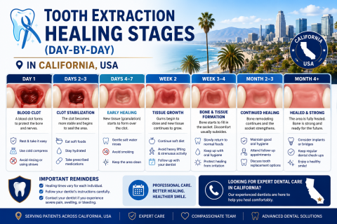
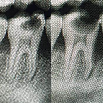
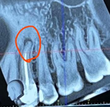
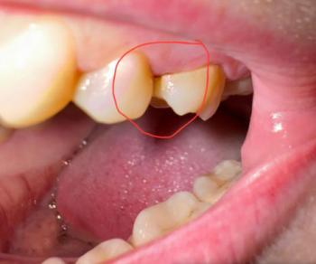
Back Tooth Decay Case Analysis: Causes, Treatment, and Healing Timeline

Severity: 

What Is Seen in This Case?

The image shows visible decay on back teeth (molars), with damaged tooth structure and discoloration. The surrounding gum tissue appears slightly irritated, which may indicate early inflammation caused by trapped bacteria.

Back teeth are more prone to decay because they are harder to clean and have deep grooves that trap food and plaque.


Most Likely Diagnosis

Based on the visual findings, the most likely conditions include:

  • Dental caries (tooth decay) on molars

Dark Buildup and Tooth Decay Case: Causes, Treatment, and Healing Timeline

Severity: 

What Is Seen in This Case?

The image shows dark buildup and damaged tooth structure on the upper teeth, with surrounding gum redness. The dark material may be severe plaque, tartar, decayed tooth structure, or infection around old dental work.

This condition usually develops over time and requires professional dental care.


Most Likely Diagnosis

Based on visual assessment, the most likely conditions include:

  • Advanced dental caries (tooth decay)

  • Heavy plaque and tartar buildup

Gum Growth Near Back Tooth: Causes, Treatment, and Healing Timeline

Severity: 

What Is Seen in This Case?

The image shows a red, swollen growth on the gum near a back tooth (molar). The surrounding tooth appears intact, but the gum tissue is inflamed and raised. This type of lesion is commonly associated with local infection, irritation, or gum overgrowth.

This condition should not be ignored, even if pain is mild.


Most Likely Diagnosis

Based on visual examination, the most likely conditions include:

  • Localized gum infection

  • Gingival abscess

Lower Front Gum Inflammation Case Analysis and Healing Timeline

Severity: 

Teeth Case – Full Analysis and Provisional Diagnosis (Zoom 100%)

This assessment is based on a single photographic image. A definitive diagnosis requires an in-person dental examination, periodontal probing, and radiographs if indicated.


1. Visual Findings (Deep Examination)

  • Teeth condition:

    • No obvious cavities or fractures visible.

Gum Inflammation and Gingivitis Case Analysis With Healing Timeline

Severity: 

Teeth Case – Full Analysis and Provisional Diagnosis (Zoom 100%)

This assessment is based on a photographic image only. A definitive diagnosis requires an in-person dental examination, periodontal probing, and radiographs when indicated.


1. Visual Findings (Deep Examination)

  • Teeth alignment and surfaces:

    • Teeth appear generally well-aligned with smooth enamel surfaces.

    • No obvious cavities, fractures, or restorations visible in this image.

Pages