Apical Infection

Pediatric Tooth Decay Case Analysis and Treatment Timeline

Severity: 

Teeth Case – Full Analysis and Provisional Diagnosis (Zoom 100%)

This assessment is based solely on a photographic image. A confirmed diagnosis requires an in-clinic dental examination, radiographs, and professional evaluation by a licensed dentist, especially for pediatric patients.


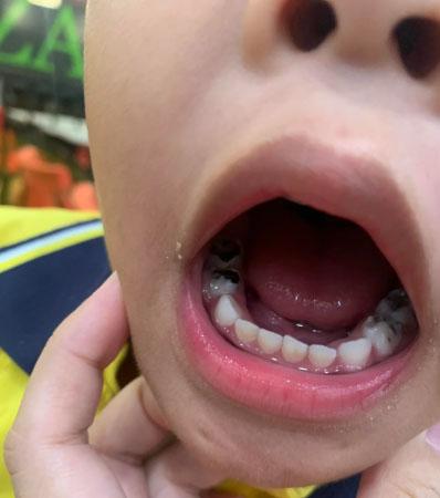
1. Visual Findings (Image-Based)

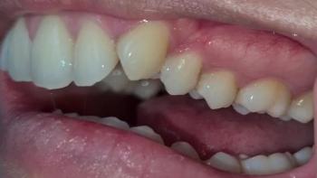
  • Lower posterior teeth show silver restorations consistent with amalgam or stainless-steel fillings.

Periapical Radiolucency Explained: Causes, Diagnosis, and Fastest Treatment for Apical Infection

Severity: 

Periapical Radiolucency Case: Apical Infection, Bone Loss, and 14-Day Healing Guide


FULL ANALYSIS (X-RAY INTERPRETATION)

1. Radiolucent Lesion at the Apex

Both Image A and Image B show a dark, well-defined radiolucent area surrounding the root apex of one of the anterior teeth. This appearance is consistent with:

  • Periapical abscess

  • Periapical cyst

  • Chronic apical granuloma

  • Chronic apical periodontitis

Pages