Gum disease

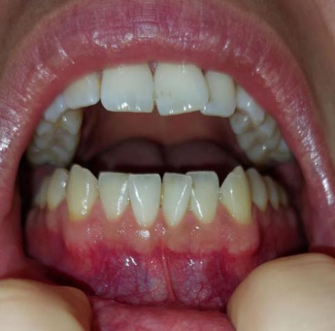
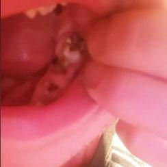
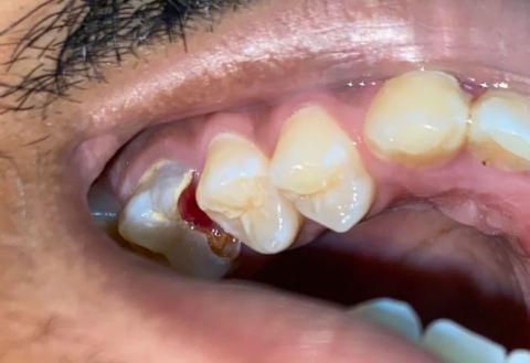
Back Tooth Decay Case Analysis: Causes, Treatment, and Healing Timeline

Severity: 

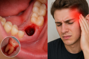
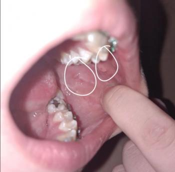
What Is Seen in This Case?

The image shows visible decay on back teeth (molars), with damaged tooth structure and discoloration. The surrounding gum tissue appears slightly irritated, which may indicate early inflammation caused by trapped bacteria.

Back teeth are more prone to decay because they are harder to clean and have deep grooves that trap food and plaque.


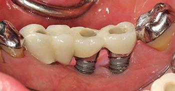
Most Likely Diagnosis

Based on the visual findings, the most likely conditions include:

  • Dental caries (tooth decay) on molars

Lower Front Gum Inflammation Case Analysis and Healing Timeline

Severity: 

Teeth Case – Full Analysis and Provisional Diagnosis (Zoom 100%)

This assessment is based on a single photographic image. A definitive diagnosis requires an in-person dental examination, periodontal probing, and radiographs if indicated.


1. Visual Findings (Deep Examination)

  • Teeth condition:

    • No obvious cavities or fractures visible.

Severe Broken and Infected Teeth Case Analysis With Low-Cost Treatment Options

Severity: 

Teeth Case – Full Analysis and Provisional Diagnosis (Zoom 100%)

Medical disclaimer:
This assessment is based on a single image and patient statement. A confirmed diagnosis requires an in-person dental examination, radiographs, and professional evaluation. Dental infections can become serious and should not be ignored.


1. Visual Findings (Deep Examination)

  • Severely broken teeth:

    • One or more teeth appear fractured down to the root level.

Teeth Misalignment with Gum Irritation - Zoomed Dental Case Analysis

Severity: 

Teeth Case – Zoom 100%

Focused Area: Upper and lower teeth alignment with surrounding gingival tissue


Full Analysis and Deep Visual Examination

This assessment is based solely on the provided image and does not replace a comprehensive dental examination, orthodontic evaluation, or dental X-rays.

Observed Findings

  • Noticeable tooth misalignment and crowding, particularly in the upper arch

  • Irregular tooth positioning creating plaque-retentive areas

Advanced Tooth Decay with Bleeding Gums - Zoomed Dental Case Analysis

Severity: 

Teeth Case – Zoom 100%

Focused Area: Posterior tooth with visible cavity and adjacent bleeding gum tissue


Full Analysis and Deep Examination

This analysis is based on the provided image only and does not replace a full clinical examination, dental probing, or radiographic assessment.

Observed Findings

  • Large cavitated lesion on the posterior tooth, with visible loss of enamel and dentin

  • Active gum bleeding adjacent to the decayed tooth

Pages