Post Root Canal Infection With Sutures Case Analysis and Treatment Guide
Severity:
Teeth Problems:
What Is Seen in This Case
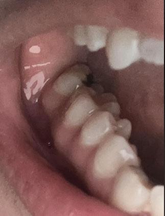
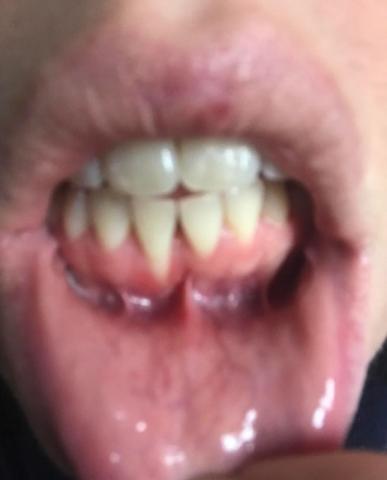
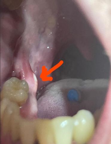
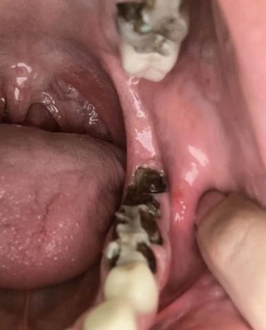
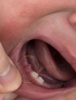
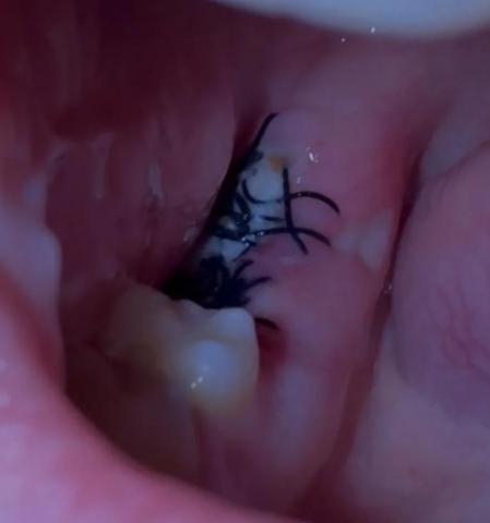
The image shows a treated tooth with sutures placed on the surrounding gum tissue. The area appears reddened and swollen, indicating recent root canal–related surgical treatment or management of an infection at the root area. The presence of sutures suggests that the infection required surgical access, drainage, or gum repair.
This is a normal appearance shortly after treatment but requires close monitoring.