Smile Again with Comfort and Confidence.
Hard Gum Lump Beside a Filled Tooth: Full Dental Analysis, Causes, and Treatment Options
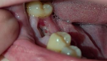
Image:
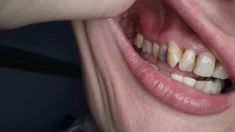
Severity:
Zoom 100% – Visual Examination Findings
Visible observations:
-
Localized firm swelling (hard lump) on the gingiva beside an upper posterior tooth
-
Surrounding gum tissue shows signs of chronic inflammation
-
Tooth involved appears discolored with an old/temporary restoration
-
Gingival margin irregular with visible periodontal pocketing
-
No obvious pus drainage seen at rest, but tissue looks chronically irritated
Client History (Highly Relevant)
-
Lump present since February (long duration)
-
Becoming harder and slightly larger
-
Vertical sides feel bone-like
-
Tender when firm pressure is applied
-
History of:
-
Severe toothache
-
Cold sensitivity progressing to constant pain
-
Temporary filling placed (June last year)
-
Diagnosed gum disease with deep pockets
-
Food trapping consistently in the area
-
Most Likely Diagnoses (Ranked)
1. Chronic Periapical Abscess with Bone Involvement (MOST LIKELY)
Why:
-
History of deep decay and unresolved tooth infection
-
Temporary filling left long-term
-
Hard swelling suggests bone reaction or chronic infection
-
Tender on pressure, not acute pain → chronic phase
Infection may be draining slowly or encapsulated, forming a firm mass.
2. Periodontal Abscess / Localized Advanced Periodontitis
Why:
-
Documented deep periodontal pocket
-
Food impaction
-
Gum disease history
-
Abscesses from periodontal origin often feel firm initially
3. Reactive Bone Growth (Exostosis / Localized Bone Response)
Why (less likely but possible):
-
Hard, bone-like consistency
-
However, tenderness and history of infection make this secondary, not primary
4. Dental Cyst (Radicular or Periodontal)
Why to consider:
-
Long-standing lesion
-
Progressive growth
-
Often painless until advanced
Requires X-ray or CBCT for confirmation.
Is the Lump Related to the Tooth with Temporary Filling?
✅ Yes – Very likely
-
Temporary fillings are not designed for long-term use
-
Bacteria may still be present in the pulp or root
-
Infection can migrate into:
-
Bone
-
Periodontal tissues
-
Surrounding gum
-
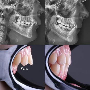
Recommended Diagnostic Process
Immediate Steps
-
Periapical X-ray or CBCT scan
-
Periodontal probing to measure pocket depth
-
Pulp vitality testing
-
Clinical palpation and percussion test
Treatment Options (Depends on Diagnosis)
If Tooth Is Restorable
-
Root Canal Treatment
-
Permanent restoration or crown
-
Periodontal deep cleaning (SRP)
If Tooth Is Not Restorable
-
Tooth extraction
-
Drainage of infection
-
Bone debridement if needed
If Periodontal Origin
-
Deep scaling and root planing
-
Local antibiotics
-
Possible periodontal surgery
Time Frame to Heal
| Phase | Expected Healing |
|---|---|
| Pain & inflammation reduction | 3–7 days |
| Soft tissue healing | 7–14 days |
| Bone healing (if involved) | 4–12 weeks |
14 days is realistic for soft tissue comfort, but bone healing takes longer.
If Left Untreated – What Will Scale Up
-
Spread of infection into jawbone
-
Facial swelling
-
Sinus involvement (upper teeth)
-
Tooth loss
-
Bone destruction
-
Systemic infection risk (rare but serious)
Professional Comments
-
A hard gum lump lasting months is NOT normal
-
Temporary fillings should never be left long-term
-
Chronic dental infections often become painless but destructive
-
Early intervention prevents surgery and bone loss
Find a Dentist Near You
Search verified dental clinics near your location:
https://cebudentalimplants.com/map-dental-clinic
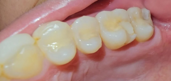
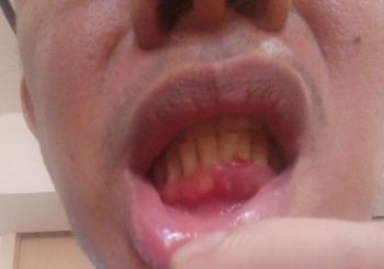
Teeth Case
 |
 |
 |
 |
 |
 |