Xray

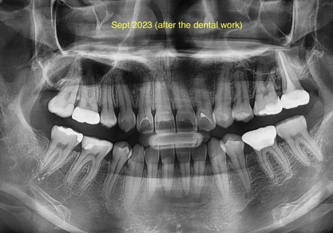
Panoramic Dental X-Ray Case Analysis After Dental Treatment
Severity: 

What Is Seen in This X-Ray

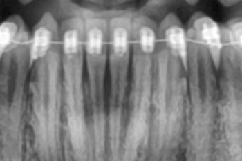
This panoramic dental X-ray (taken after dental work in September 2023) shows multiple restored teeth, including fillings and treated posterior teeth. The roots and surrounding bone are visible across the upper and lower jaws, allowing evaluation of healing, hidden infection, and overall oral health.

Some areas show treated teeth with restorations, while other regions require close monitoring for possible residual or recurrent infection.
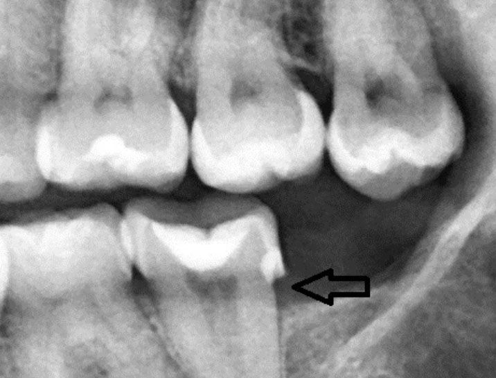
Interproximal Tooth Decay Seen on Dental X-ray - Zoomed Case Analysis

Severity: 
Teeth Problems: 

Teeth Case – Zoom 100% (Dental X-ray)

Focused Area: Posterior teeth (molar region) with interproximal defect indicated by arrow


Full Analysis and Deep Radiographic Examination

This assessment is based on the provided dental X-ray image only and does not replace a complete clinical examination, periodontal probing, or additional radiographs if required.

Radiographic Findings

  • Radiolucent area between posterior teeth, consistent with interproximal dental caries

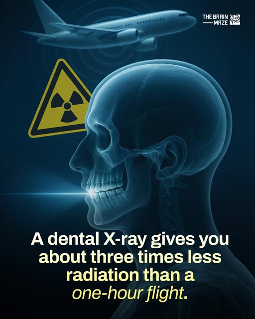
Your Flight vs. Your X-Ray: Understanding Radiation Exposure for Travelers

Topics: 

Clearing the Air: A Traveler’s Guide to Dental X-Ray Safety in the Philippines

For the modern traveler, a trip to the Philippines is an adventure of stunning landscapes, vibrant culture, and—sometimes—unexpected events. While you meticulously plan your itinerary through Cebu’s waterfalls or Palawan’s lagoons, one thing you likely don't plan for is a dental emergency. If you find yourself in a clinic needing an X-ray, a common concern arises: "How much radiation am I being exposed to?"

Pages