


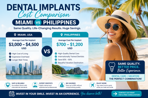
Dental Implants Cost Miami vs Philippines: An Expert Perspective from Miami (2026 Guide)
Topics:
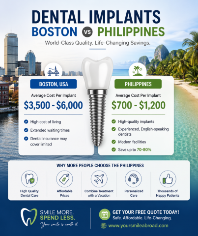
As a dental implant expert practicing in Miami, I regularly meet patients who are weighing one key question: Should I invest in dental implants locally, or travel abroad to save money?
The comparison between Miami and the Philippines is one of the most common discussions today—especially among patients seeking high-quality results at a more affordable cost.
This guide provides a clinical and practical breakdown of pricing, quality, risks, and long-term value—so you can make a confident and informed decisione.