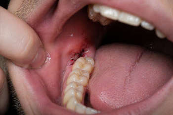
Deep Cavity on Back Molar With Early Gum Infection
Severity:
Teeth Problems:
Deep Cavity on Back Molar With Early Gum Infection Case Analysis
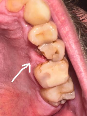
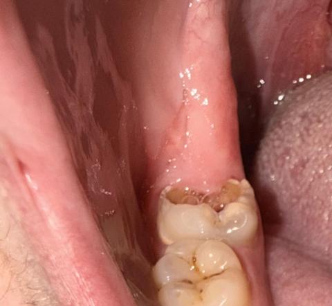
What Is Seen in This Case
The image shows a back molar with a deep cavity on the chewing surface, where the enamel has broken down and darker material is visible inside the tooth. The gum tissue beside the tooth looks irritated and slightly inflamed, suggesting early gum infection next to an actively decaying molar. The tongue is close to the area, which may increase discomfort.